FIGURE 1. Cladogram representing the Epigondolella clade of Mazza et al. (2012b), illustrating the phylogenetic relationships between the Upper Carnian-Lower Norian (Upper Triassic) carnepigondolellids and epigondolellids and their main evolutionary trends. Only the species analysed in this work are figured. The specimens of E. vialovi, E. quadrata, E. uniformis, and E. triangularis are from Mazza and Martínez-Pérez (2015). Vertical bars beside the specimens indicate the platform (P)/blade (B) length ratio; white circles mark the cusp. All the specimens are at the same scale.
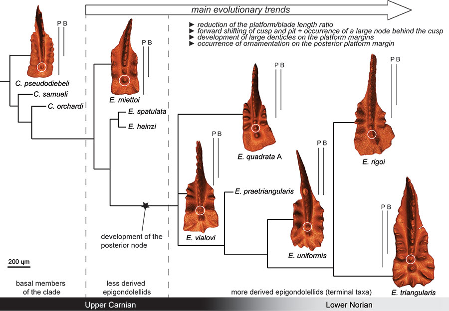
FIGURE 2. X-ray Synchrotron microtomography of Epigondolella quadrata specimen A. Three possible sections that can be obtained with the X-ray synchrotron microscopy are shown. 1, 3D model of the specimen: 2, Longitudinal section; 3, Horizontal section; 4, Cross section. Scale bar equals 200 µm.
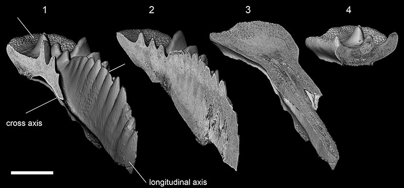
FIGURE 3. X-ray synchrotron microtomographic sections compared to SEM photos of artificially fractured conodont specimens. 1, Tomographic section of Epigondolella quadrata specimen A; 2-4, SEM photos of an artificially fractured specimen of Carnepigondolella carpathica (sample NA16). Both the specimens are sectioned in correspondence to the cusp; 5-6, Comparison between a tomographic section (5) and an artificially fractured (6) specimen of E. uniformis from the same sample (NA42). Legend: cu, cusp; wm, white matter; hp, hypocalcification; gl, growth lines.
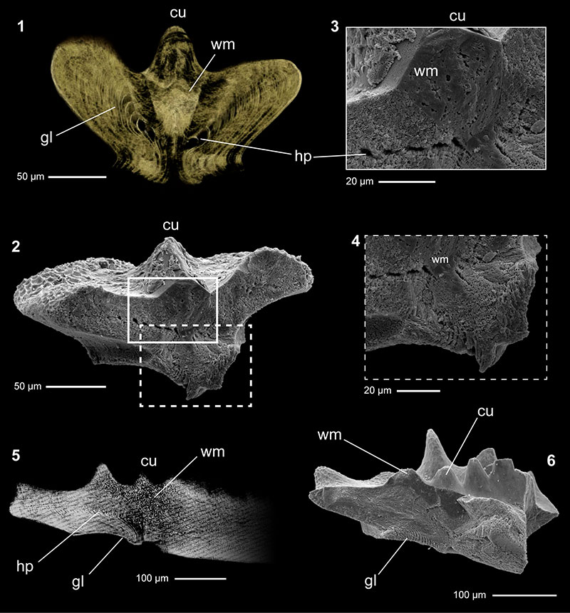
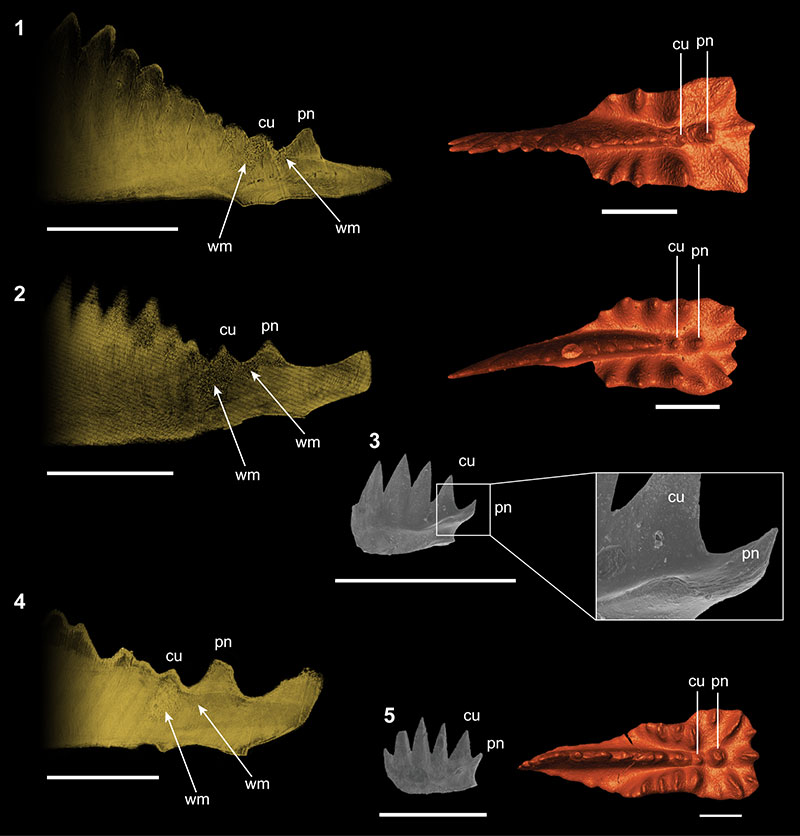
FIGURE 4. Microtomographic longitudinal sections focused on the posterior platform of Epigondolella quadrata and E. rigoi, aimed to show the ontogenesis of the posterior node growing behind the cusp. For each section a 3D model of the correspondent conodont is provided. Beside the sections, the outlines of selected growth lines are reported, in order to evidence some visible growth stages and show the different ontogenetic processes of E. quadrata and E. rigoi. Arrows and letters (a, b, c) mark the growth lines considered to draw the stages. 1, E. quadrata A; 2, juvenile specimen of E. quadrata from sample NA60 (from Mazza and Martínez-Pérez, 2015; repository number Micro-Unimi no. 2001); 3, E. quadrata B; 4, E. quadrata C; 5, E. rigoi A; 6, juvenile specimen of E. rigoi from sample NA68 (from Mazza and Martínez-Pérez, 2015; repository number Micro-Unimi no. 2003), showing that the posterior node is already occurring; 7, E. rigoi B. Scale bars equal 200 µm. Legend: cu, cusp; pn, posterior node.
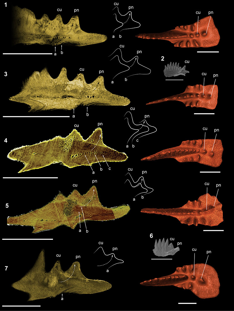
FIGURE 5. Microtomographic longitudinal sections focused on the posterior platform of Epigondolella vialovi, E. uniformis, and E. triangularis aimed to show the ontogenesis of the posterior node growing behind the cusp. For each section a 3D model of the correspondent conodont is provided. 1, E. vialovi ; 2, E. uniformis ; 3, juvenile specimen of E. uniformis from sample NA43 (from Mazza and Martínez-Pérez, 2015; repository number Micro-Unimi no. 2005); 4, E. triangularis ; 5, juvenile specimen of E. triangularis from sample NA43 (from Mazza and Martínez-Pérez, 2015; repository number Micro-Unimi no. 2004). Scale bars equal 200 µm. Legend: cu, cusp; pn, posterior node; wm, white matter.