FIGURE 1. 1, Location map and 2, log of the Wadi Al Fayn section (by courtesy of Dujoncquoy, 2011). Star: Type-level of Draconisella mortoni sp. nov., 1) Wadi Nakhr (23°10'26.7"N 57°12'00.4"E), 2) Wadi Kamah (23°06'55.7"N 57°33'07.3"E), 3) Wadi Mu Aydin (23°04'40.1"N 57°29'39.3"E), 4) Wadi Tanuf (22°58'35.3"N 57°40'05.0"E), and 5) Wadi Al Fayn (23°04'39.63"N 57°47'12.55"E). Color code for the main facies: marls and mudstones in blue, bioclastic wackestones in green, oolitic grainstones in yellow, bioclastic and oolitic grainstones in orange, rudist or oncoid floatstones in pink or red.
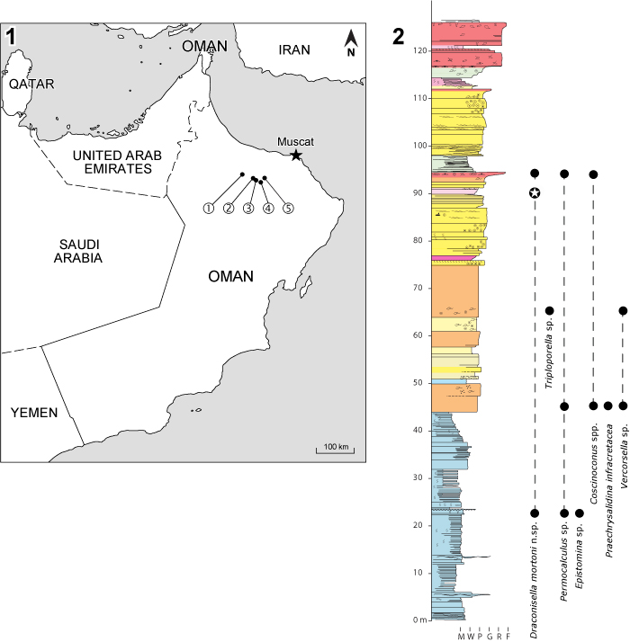
FIGURE 2. Microfossils, “calcareous” algae and foraminifera. 1-2, Deloffrella hauteriviana (Masse, 1999, non 1976): 1, subaxial and oblique sections, Wadi Kamah, sample no. 7 (= Granier, 2013, figure 2.13), 2, subaxial sections,Wadi Mu Aydin, sample no. base-1 (= Granier, 2013, figure 2.3); 3-4, Holosporella sugdeni (Elliott, 1957) Granier, 2017, obliques sections, Wadi Kamah, sample no. 33bis; 5, Praechrysalidina infracretacea Luperto Sinni, 1979, axial section, Wadi Nakhr, sample no. 3; 6-8,Falsolikanella danilovae (Radoičić ex Barattolo, 1978), Wadi Tanuf: 6, oblique section, sample no. 30, 7, subtransverse section, sample no. 54, 8, oblique section, sample no. 31; 9-10, Mayncina sp., Wadi-Kamah: 9, tangential oblique section, sample no. 35, 10, oblique section, sample no. 16; 11-12, Choffatella decipiens Schlumberger, 1905, Wadi Kamah: 11, oblique section, sample no. 34, 12, subaxial section, sample no. 14; 13-16, Coscinoconus sp., Wadi Al Fayn, oblique sections, sample no. 33; 17, Epistomina sp., subaxial section, Wadi Kamah, sample no. 34. Scale bar (for all photos) equals 250 µm.
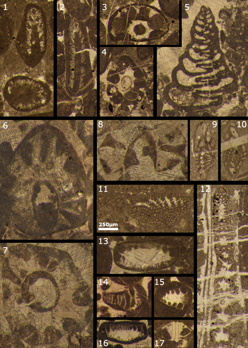
FIGURE 3. Draconisella mortoni sp. nov.: 1-3, Wadi Mu Aydin, sample “base”; 4, holotype, Wadi Al Fayn, sample no. 31; 5-13, Wadi Kamah; 5-6, sample no. 13; 7-9, sample no. 14; 10, sample 31; 11-12, sample no. 32. 1, 4, 9 and 11 are subaxial sections (the main axis is the open pore on the lower side); 7 and 10 are tangential sections (they do not reach the main axis); 2-3, 5-6, 8 and 12 are oblique sections. Scale bar (for all photos) equals 100 µm.
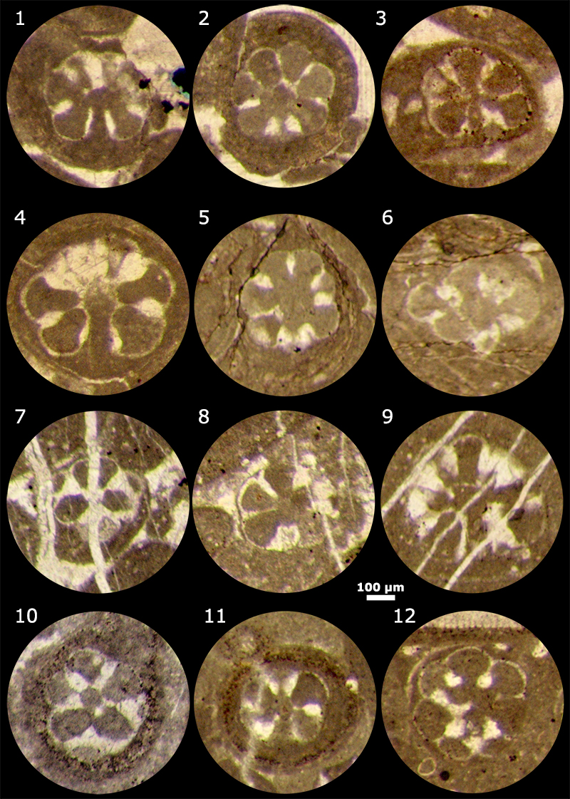
FIGURE 4. Mizzia velebitana Schubert, 1909: 1, three articles partly encrusted by bryozoans and still connected, sample no. 692, Collection J. von Pia, “New Mexico, SW of Carlsbad”; 2-4, sample no. CXIV, “Mizziendolomit, Oberstes Karbon. Paklenica, Velebit Geb., Dalmatien. Coll. Schubert”. 2, tangential section of an article, thin section CXIV-1 (Pia, 1920, plate I, figure 13); 3, transverse section of an article, thin section CXIV-3 (Pia, 1920, plate I, figure 21); 4, axial section of an article, neotype herein defined, thin section CXIV-3 (Pia, 1920, plate I, figure 20). Photo 1: Scale bar equals 500 µm; photos 2-4: Scale bar equals 250 µm.
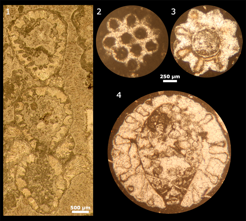
FIGURE 5. Draconisella genoti Granier and Michaud, 1989: 1, deep tangential section of an article (= Granier and Michaud, 1989, plate 1, figure 2); 2, deep tangential section of an article (= Granier and Michaud, 1989, plate 1, figure 3); 3-10, deep tangential sections of loose articles; 11, holotype, oblique section of an article with axial cavity visible (= Granier and Michaud, 1989, plate 1, figure 5); 12, deep tangential section section of an article (= Granier and Michaud, 1989, plate 1, figure 6); 1-4, sample no. MX 84 57; 5-6, sample no. MX 84 63; 7-9, sample no. MX 85 495; 10-12, sample no. MX 85 371. Scale bar (for all photos) equals 250 µm.
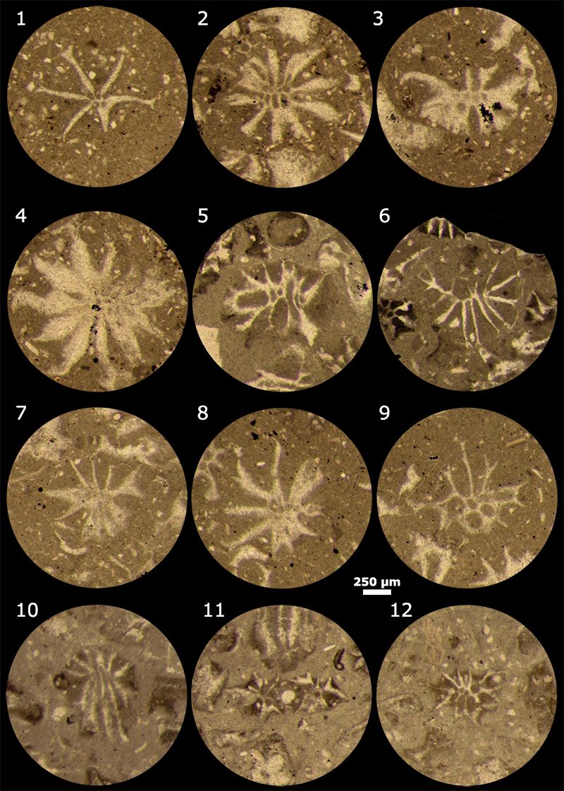
FIGURE 6. 3D “Blender” model for Mizzia-like alga. The main axis is regularly inflated and the laterals are thin in their proximal part. Click on image to run or download animation.
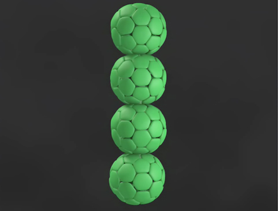
FIGURE 7. 3D “Blender” model for Draconisella-like alga. The main axis is rather thin, and the laterals are inflated in their proximal part. Click on image to run or download animation.
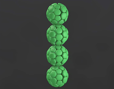
FIGURE 8. Rajkaella iailaensis Maslov ex Dragastan and Bucur, 1988, non 1965. 1-2 and 4-6, various oblique to tangential oblique sections through a verticil; 5, tangential section of a lateral at its distal end, through the secondary laterals. 1-3, Berriasian, Corbières (France), Collection Jaffrezo; 4-6, Berriasian, French Jura (France). R1: primary lateral; R2: secondary laterals. Scale bar (for all photos) equals 250 µm.
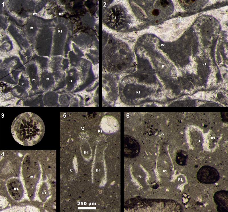
FIGURE 9. The five morphotypes of living Dasycladalean thalli. 1, cylindrical thallus (Batophora oerstedi Agardh, 1854, excerpt from Berger, 2006, Fig. 15); 2, claviform, club-shaped thallus (Bornetella oligospora Solms-Laubach, 1893, excerpt from Berger, 2006, Fig. 33); 3-4, articulated, moniliform thalli, branched (Cymopolia barbata Lamouroux, 1816, excerpt from Berger, 2006, Fig. 64) and not (Cymopolia vanbosseae Solms-Laubach, 1893, excerpt from Berger, 2006, Fig. 72); 5, sphaerical, ball-shaped thallus (Bornetella sphaerica (Zanardini, 1878), excerpt from Berger, 2006, Fig. 43); 6-7, umbelliform thalli (6: Acetabularia kilneri Agardh, 1886, excerpt from Berger, 2006, Fig. 166; 7: Acetabularia schenckii Möbius, 1899, excerpt from Berger, 2006, Fig. 107). Scale bars 1-2 equals 2.5 mm, 3 equals 10 mm, 4-5 equals 1 mm, 6-7 equals 5 mm.
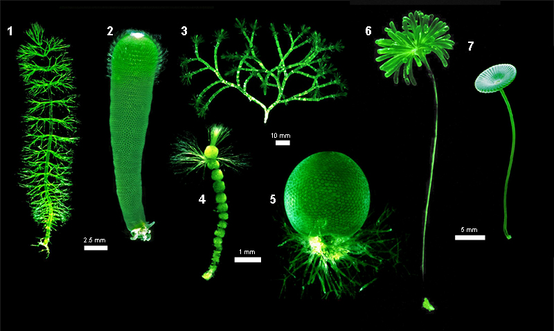
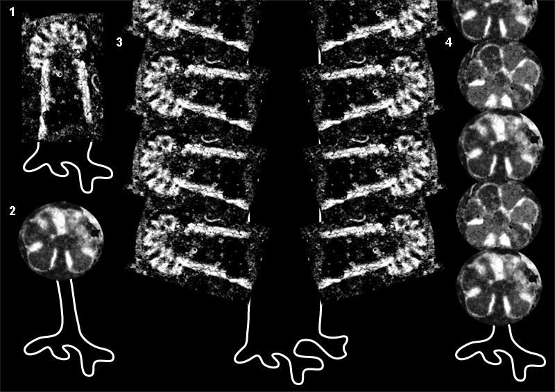
FIGURE 10. 1, the original interpretation of Koptedagaria iailaensis Maslov, 1965, nom. nud., which has proved to be wrong (Granier, 1990); 2, the inarticulate-sphaerical hypothetical reconstruction of the new Omani alga, which is not sustainable; 3, the revised interpretation of Rajkaella iailaensis Maslov ex Dragastan and Bucur, 1988, non 1965, with a large cylindrical thallus bearing verticils of rather large laterals (as documented in Figure 8); 4, the articulate-moniliform reconstruction of Draconisella mortoni sp. nov., which is finally adopted in this study.