FIGURE 1. Location of the sampling sites in the northeastern Indian Ocean. The surface current (thick arrows) is modified from Peng et al. (2015); the deep circulation (thin arrows) is modified from Kawagata et al. (2006); the isohaline showing surface salinity during June (mean of 93 years) is modified from Gupta et al. (2002). Abbreviations are as follows: AABW, Antarctic Bottom Water; CPDW, Circumpolar Deep Water; NIHSIW, North Indian High-salinity Intermediate Water; NMC, Northeast Monsoon Current; SMC, Southwest Monsoon Current.
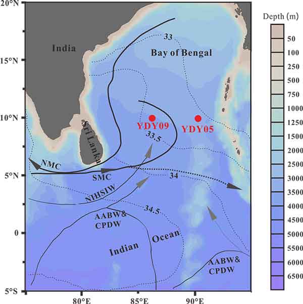
FIGURE 2. Morphological terminology under a transmitted light microscope. 1-2, for gate and girdle; 3-8, for different pylonioid systems.
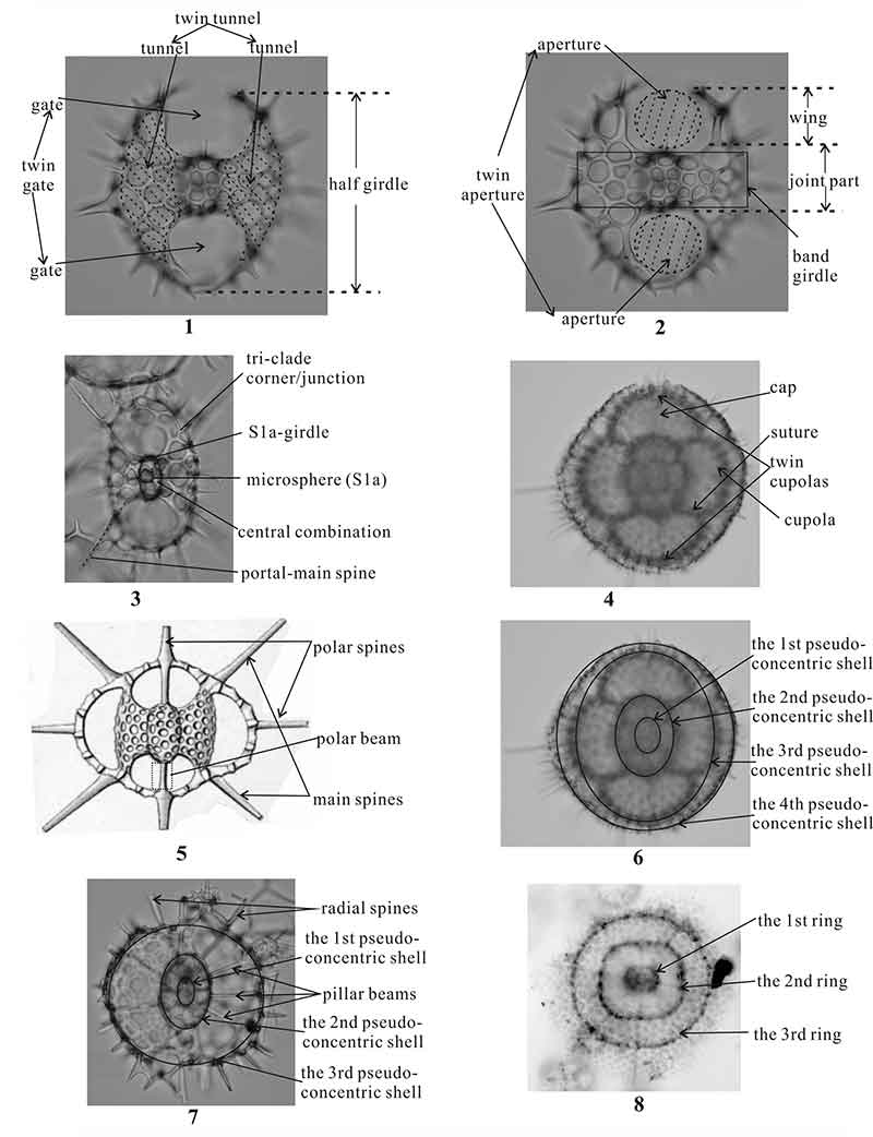
FIGURE 3. The absolute and relative orientations of specimens under the pylonioid system. The symbol “*” indicates Type 2 Relative Cartesian Coordinates.
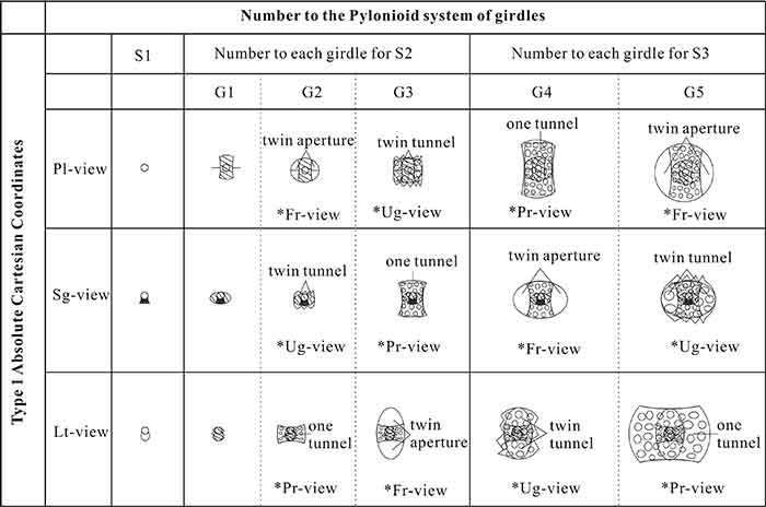
FIGURE 4. The diagrams of the absolute Cartesian coordinate (Type 1) and the relative Cartesian coordinate (Type 2). 1, Type 1; 2, Type 2.
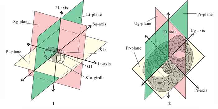
FIGURE 5. The structure of central combination in genera Tetrapyle (1-3), Larcopyle (4) and Circodiscus (5). 1, Sg-view at Type 1, Fr-view at Type 2; 2, Pl-view at Type 1, Fr-view at Type 2; 3, Lt-view at Type 1, Pr-view at Type 2. G1, G2, S1a, and S1a-girdle are the morphological terminology, of which the definitions have been shown in Table 1.
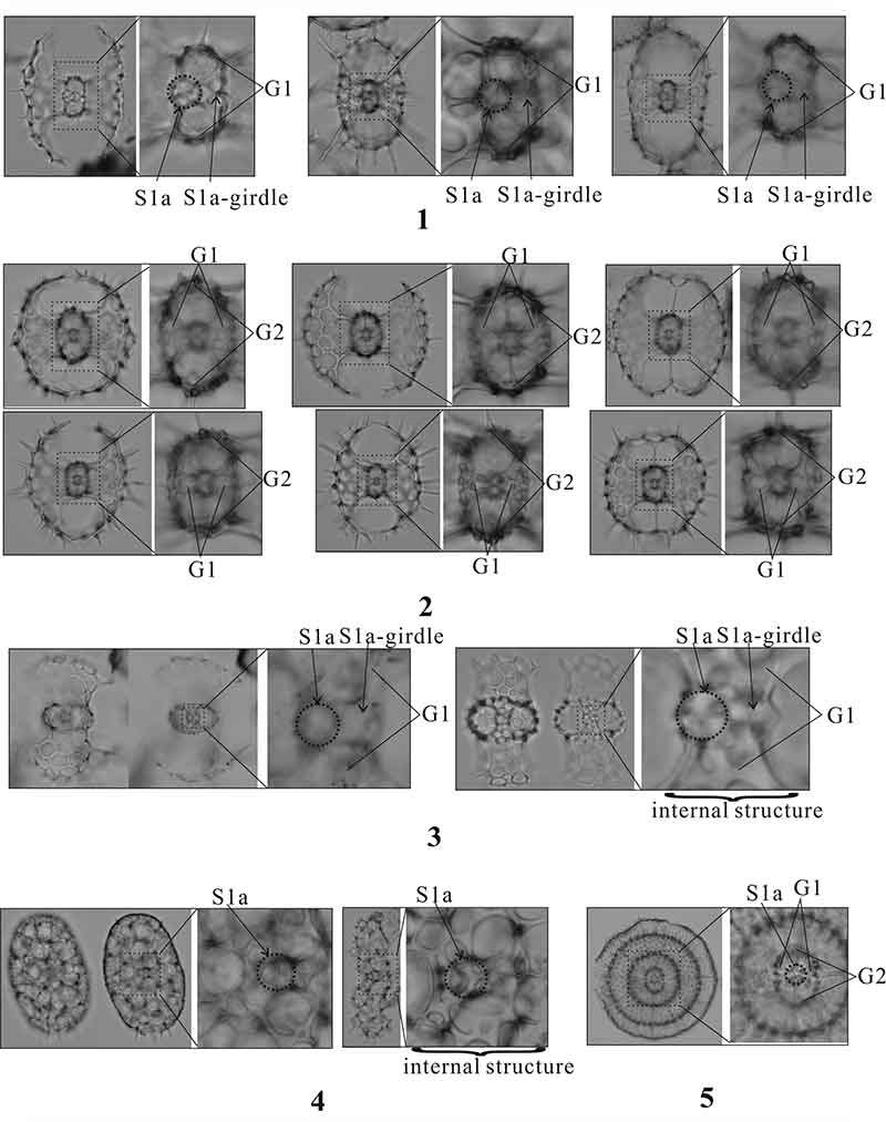
FIGURE 6. Photographs of the encountered morphotypes of Tetrapyle octacantha Müller 1859 sensu stricto. All are Pl-view at Type 1, Fr-view at Type 2. Scale bar equals 0.1 mm. The arrow marks indicate the major portal-spines. All specimens are from YDY05-01.
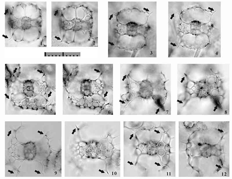
FIGURE 7. The typical Tetrapyle octacantha specimens from the surface water from the southern Villefranche-sur-Mer. 1 and 6, Ug-view at Type 2; 2-3 and 8, Pr-view at Type 2; 4-7, Fr-view at Type 2. Scale bar equals 0.1 mm. The arrow marks indicate the major portal-spines.
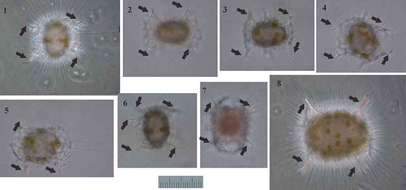
FIGURE 8. Photographs of the encountered morphotypes of Tetrapyle circularis Haeckel, 1887. All are Pl-view at Type 1, Fr-view at Type 2. Scale bar equals 0.1 mm. All specimens are from YDY05-01.
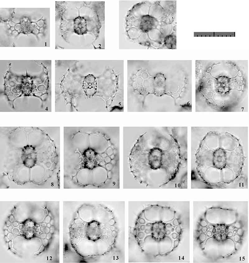
FIGURE 9. Photographs of the encountered morphotypes of Tetrapyle circularis Haeckel, 1887. All are Pl-view at Type 1, Fr-view at Type 2. Scale bar equals 0.1 mm. All specimens are from YDY05-01.
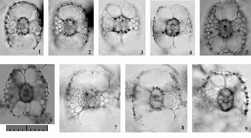
FIGURE 10. Photographs of the encountered morphotypes of Tetrapyle fruticosa (Tan and Chen, 1990) new combination. All are Pl-view at Type 1, Fr-view at Type 2. Scale bar equals 0.1 mm. All specimens are from YDY05-01.
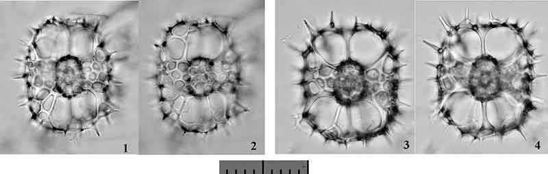
FIGURE 11. Photographs of the encountered morphotypes of Tetrapyle spp. (juvenile form). 1-2 and 9, Lt-view at Type 1, Fr-view at Type 2; 3-5, 7-8, 11-12, Sg-view at Type 1, Fr-view at Type 2; 6 and 10, Pl-view at Type 1, Fr-view at Type 2; 13-14, Pl-view at Type 1, Ug-view at Type 2; 15-16, Sg-view at Type 1, Fr-view at Type 2; 17-18, Sg-view at Type 1, Ug-view at Type 2. Scale bar equals 0.1 mm. All specimens are from YDY05-01.
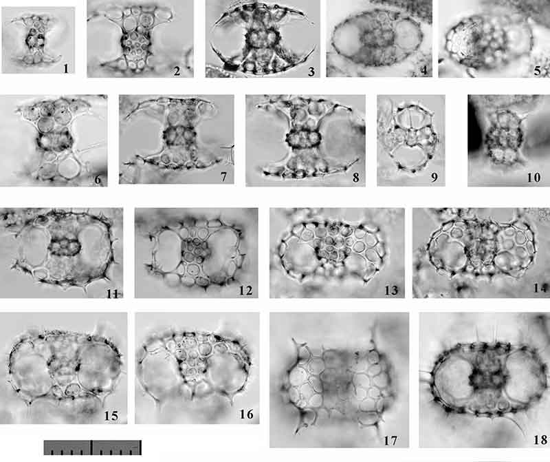
FIGURE 12. Photographs of the encountered morphotypes of Larcospira quadrangula Haeckel, 1887 sensu stricto. All are Pl-view at Type 1. Scale bar equals 0.1 mm. All specimens are from YDY05-01.
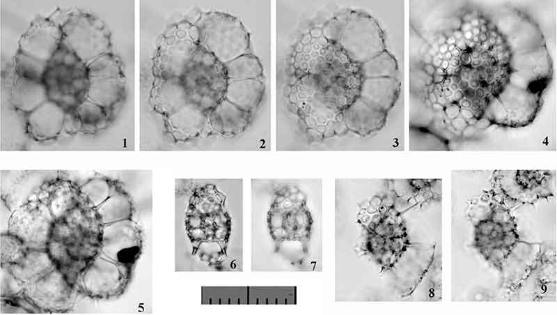
FIGURE 13. Photographs of the encountered morphotypes of Larcospira teres n. sp. All are Pl-view at Type 1. Scale bar equals 0.1 mm. All specimens are from YDY05-01.
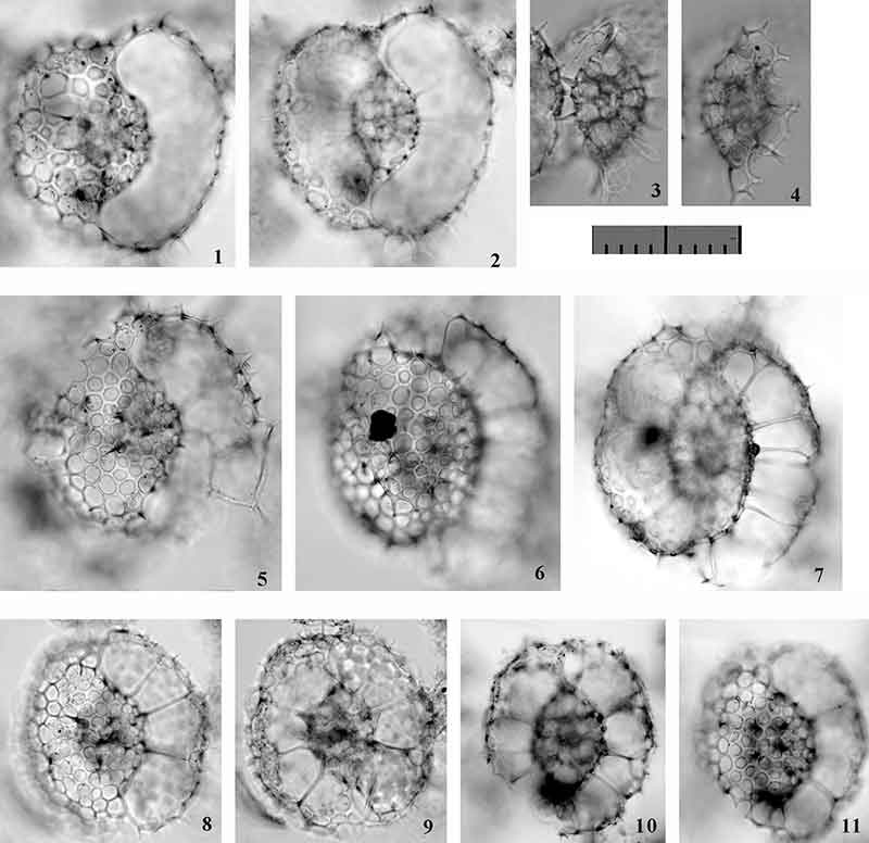
FIGURE 14. Photographs of the encountered morphotypes of Larcospira tetragonicentrum n. sp. All are Pl-view at Type 1. Scale bar equals 0.1 mm. All specimens are from YDY05-01.
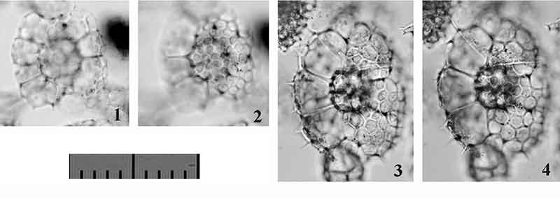
FIGURE 15. Photographs of the encountered morphotypes of Pylodiscus spinulosus (Chen and Tan, 1989). All are Fr-view at Type 2. Scale bar equals 0.1 mm. All specimens are from YDY05-01.
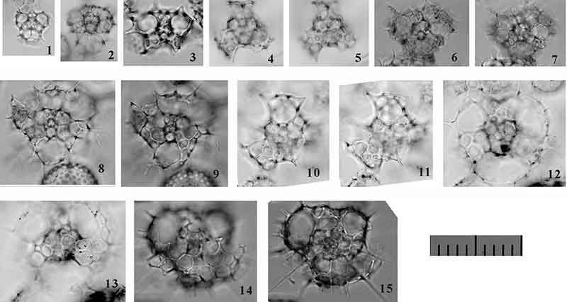
FIGURE 16. Photographs of the encountered morphotypes of Larcopyle cervicornis (Haeckel, 1887) sensu stricto. All are Pl-view at Type 1. Scale bar equals 0.1 mm. All specimens are from YDY05-01.
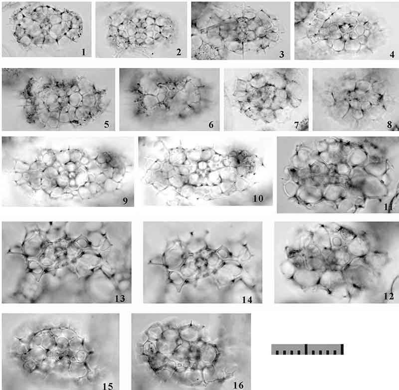
FIGURE 17. The topotypes from the H.M.S. Challenger station 271. 1-6, Larcopyle cervicornis (Haeckel, 1887); 7-12, Larcopyle buetschlii buetschlii Dreyer, 1889. All are Pl-view at Type1. Scale bar equals 0.1 mm. All specimens are from H.M.S. Challenger Station 271 sample
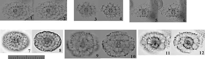
FIGURE 18. Photographs of the encountered morphotypes of Larcopyle. 1-6, Larcopyle variabile (Dreyer, 1889) sensu emend.; 7-12, Larcopyle molle n. sp.; 13-18, Larcopyle eccentricanoides n. sp.; 19-22, Larcopyle pulchella n. sp. All are Fr-view at Type 2. Scale bar equals 0.1 mm. All specimens are from YDY05-01.
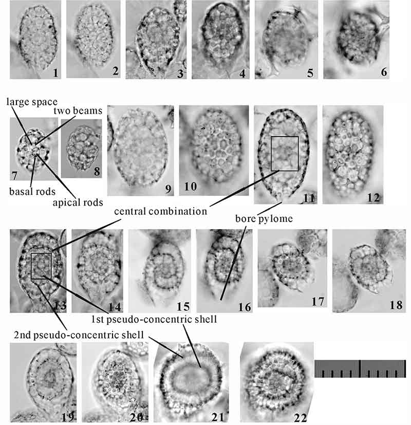
FIGURE 19. Photographs of the encountered morphotypes of Larcopyle buetschlii buetschlii Dreyer, 1889, sensu stricto. All are Fr-view at Type 2. Scale bar equals 0.1 mm. All specimens are from YDY05-01.
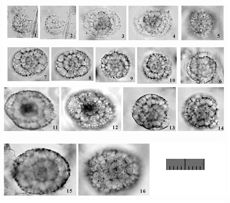
FIGURE 20. Photographs of the encountered morphotypes of Larcopyle buetschlii chenmuhongi n. subsp. All are Fr-view at Type 2. Scale bar equals 0.1 mm. All specimens are from YDY05-01.
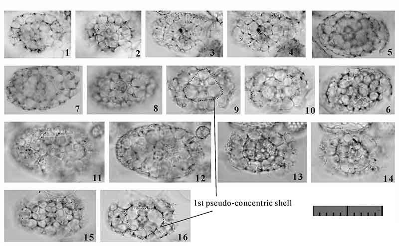
FIGURE 21. Photographs of the encountered morphotypes of Larcopyle buetschlii orion n. subsp. All are Fr-view at Type 2. Scale bar equals 0.1 mm. All specimens are from YDY05-01.
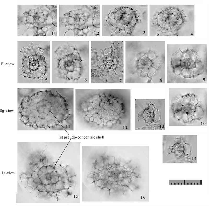
FIGURE 22. Photographs of the encountered morphotypes of Sphaeropylolena laxa n. sp. All are Fr-view at Type 2. Scale bar equals 0.1 mm. All specimens are from YDY05-01.

FIGURE 23. Photographs of the encountered morphotypes of Sphaeropylolena tenellispinosa n. sp. All are Fr-view at Type 2. Scale bar equals 0.1 mm. All specimens are from YDY05-01.
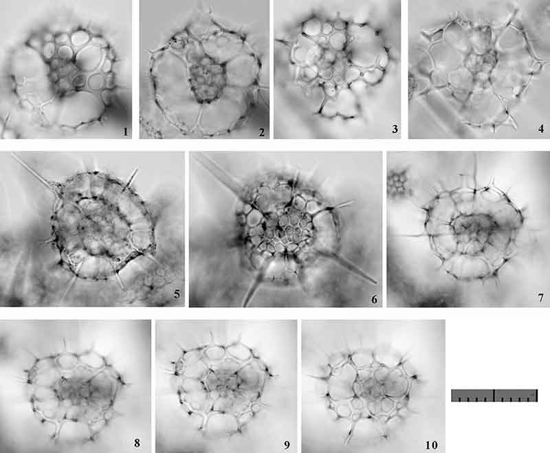
FIGURE 24. Photographs of the encountered morphotypes of Phorticium multispinum Popofsky, 1912 sensu emend. (1-3) and Phorticium itakii n. sp. (4-6). All are Pl-view at Type 1, Fr-view at Type 2. Scale bar equala 0.1 mm. All specimens are from YDY05-01.
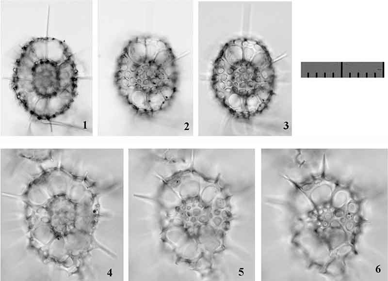
FIGURE 25. Photographs of the encountered morphotypes of Phorticium polycladum Tan and Tchang, 1976. 1-10, Sg-view at Type 1; 11-14, Pl-view at Type 1. Scale bar equals 0.1 mm. All specimens are from YDY05-01.
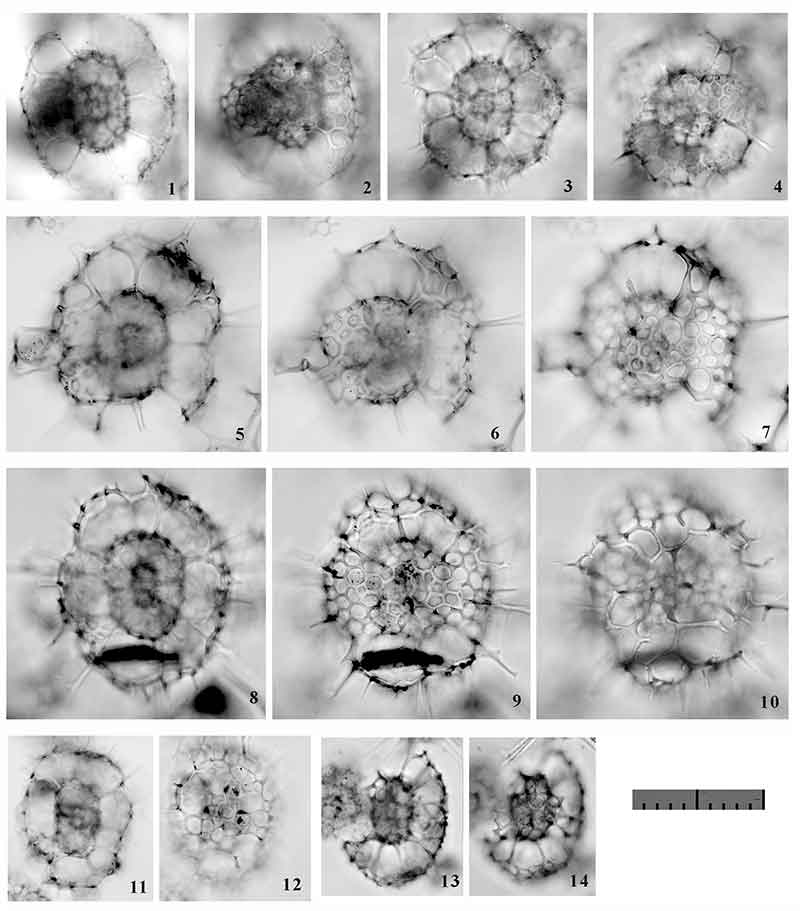
FIGURE 26. Photographs of the encountered morphotypes of Phorticium scitulum n. sp. 1-23, Sg-view at Type 1. Scale bar equals 0.1 mm. All specimens are from YDY05-01.
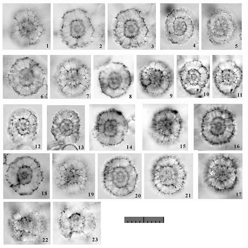
FIGURE 27. Photographs of the encountered morphotypes of Sphaerolarnacillium cochleatum gen. et n. sp. All are Fr-view at Type 2. Scale bar equals 0.1 mm. All specimens are from YDY05-01.
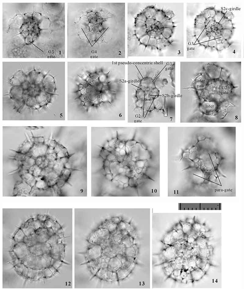
FIGURE 28. Photographs of the encountered morphotypes of Sphaerolarnacillium exactum n. sp. All are Fr-view at Type 2. Scale bar equala 0.1 mm. All specimens are from YDY05-01.
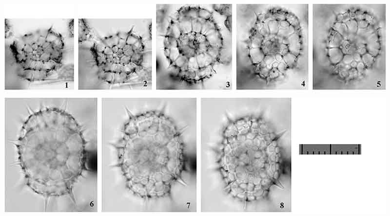
FIGURE 29. Photographs of the encountered morphotypes of Sphaerolarnacillium tanzhiyuani n. sp. All are Fr-view at Type 2. Scale bar equals 0.1 mm. All specimens are from YDY05-01.
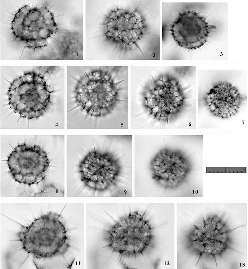
FIGURE 30. Photographs of the encountered morphotypes of Qiuripylolena. 1-10, Qiuripylolena chikuchik n. sp.; 11-22, Qiuripylolena pompon n. sp.; 23-34, Qiuripylolena ? multiconcentica n. sp. All are Sg-view at Type 1. Scale bar equals 0.1 mm. All specimens are from YDY05-01.
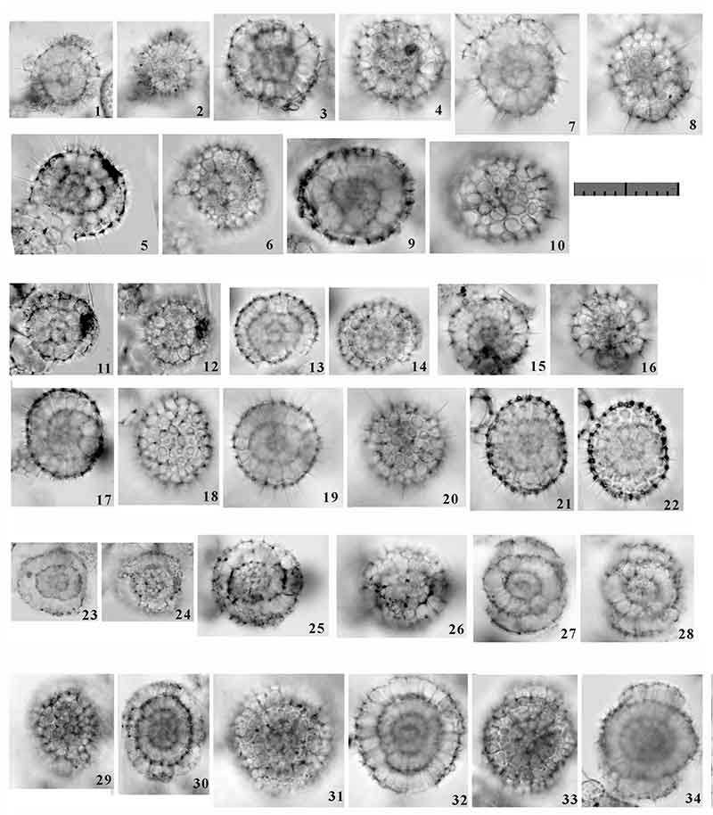
FIGURE 31. Photographs of the encountered morphotypes of Circodiscus. 1-2, Circodiscus amphitrites (Ehrenberg, 1854b); 3-4, Circodiscus biorbiculus n. sp.; 5-14, Circodiscus microporus (Stöhr, 1880); and 15-16, Circodiscus pseudomicroporus n. sp. All are Sg-view at Type1, Fr-view at Type 2. Scale bar equals 0.1 mm. All specimens are from YDY05-01.
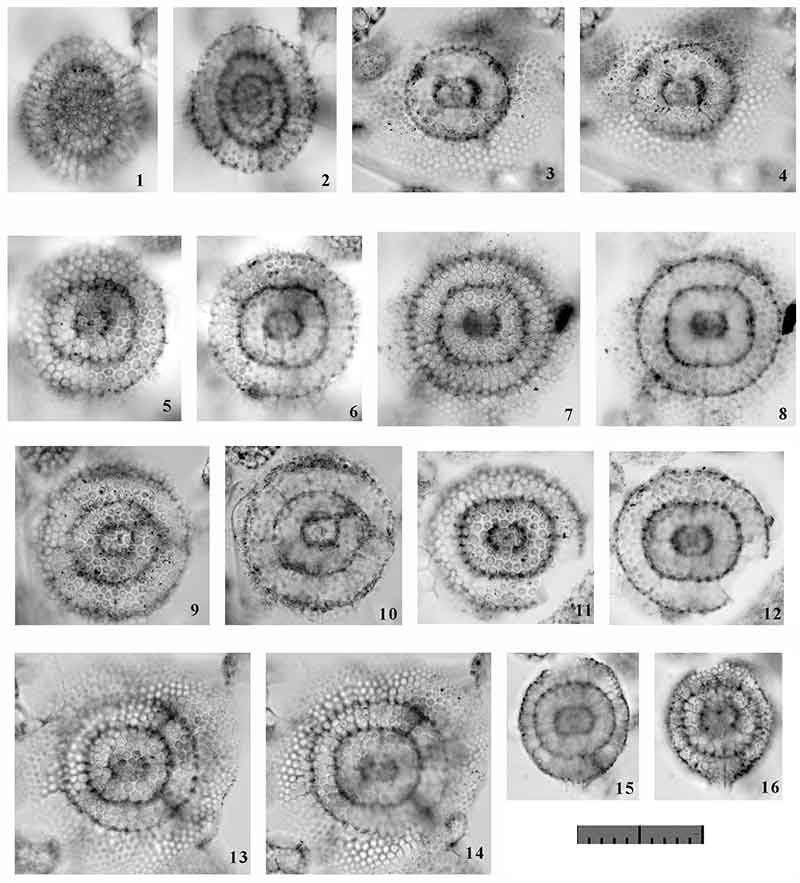
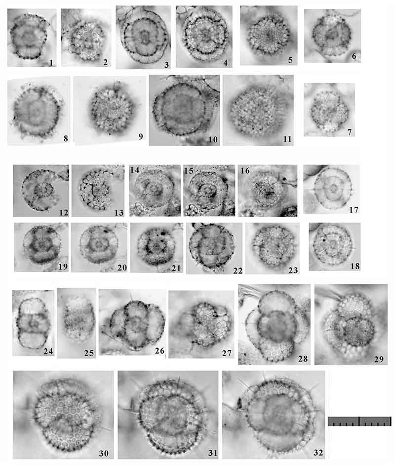
FIGURE 32. Photographs of the encountered morphotypes of Tholomura. 1-11, Tholomura hexonia (Haeckel, 1887); 12-23, Tholomura pilula n. sp.; 24-32, Tholomura polystyla (Chen, 1987). 1-16, 19-23, 26-32, Pl-view at Type 1; 17-18, 24-25, Lt-view at Type 1. Scale bar equals 0.1 mm. All specimens are from YDY05-01.