FIGURE 1. Geographic location and extension of the outcrops of the Cañadón Calcáreo Formation (represented in red) and the Cañadón Asfalto Formation (represented in dark gray) in the Chubut province, Patagonia, Argentina. The white spots on the outcrops of the Cañadón Calcáreo Formation in the detailed map on the bottom right represent the main fish localities, which are precisely indicated in Figure 2.
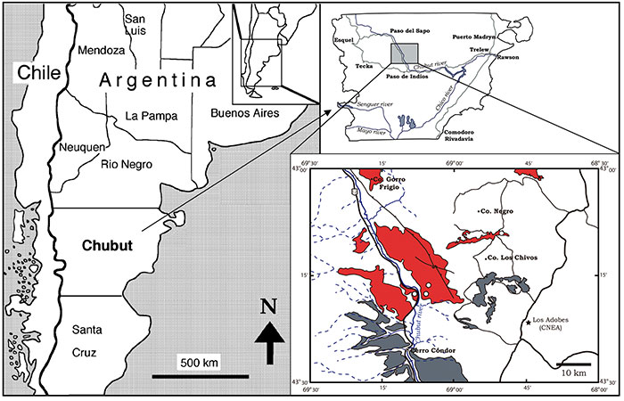
FIGURE 2. Geographic location of the fish localities: 1, Puesto Almada; 2, Punta Biotita; 3. Estancia Limonao; 4, Cañadón Los Chivos 1; 5, Cañadón Los Chivos 2; 6, Cañadón Las Minas; 7, La Manea. Geographic information was drawn on a Google earth image (© 2012 Digital Globe).

FIGURE 3. Relative abundance of the main fish groups represented in the Cañadón Calcáreo Formation. 3.1, General taxonomic composition in four of the different localities: Puesto Almada: n= 131; Estancia Limonao: n=44; La Manea: n=125; Cañadón Los Chivos 1: n=11 (see geographic distribution of these localities in Figure 2). 3.2, Detailed taxonomic composition present in the different fish-bearing layers (L1 to L7 from the top to the base of the sequence) of the La Manea locality: Layer 1: n=9; layer 3: n=18; layer 4: n=11; layer 5: n= 8; layer 6: n=11; layer 7: n=7.
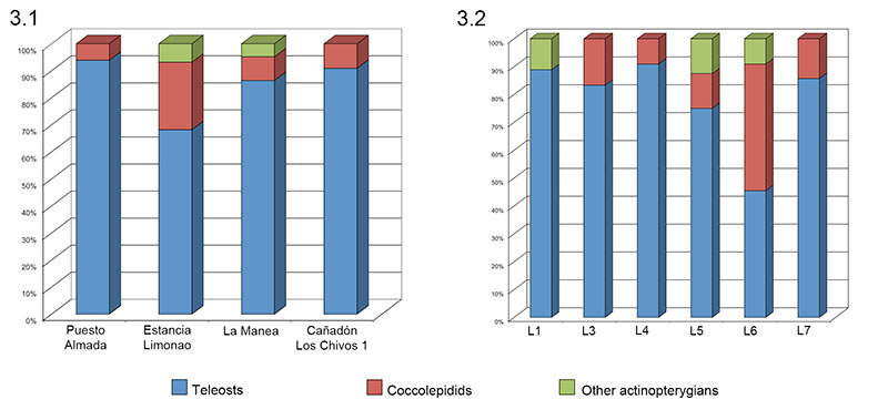
FIGURE 4. Photographs of two type specimens of †Condorlepis groeberi (Bordas, 1943) n. comb. 4.1, MACN 14434 (lectotype); 4.2, MACN 14433 (paralectotype). Scale bars equals 2 cm.
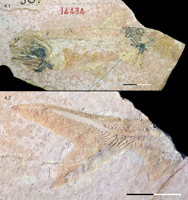
FIGURE 5. Photograph of the specimen MPEF-PV 1731A of †Condorlepis groeberi. Scale bar equals 2 cm.
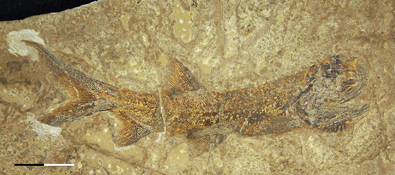
FIGURE 6. Photograph of the skull of †Condorlepis groeberi in MACN 14434 (lectotype). Scale bar equals 5 mm.

FIGURE 7. Line drawing of the skull of †Condorlepis groeberi in MPEF-PV 1731B.
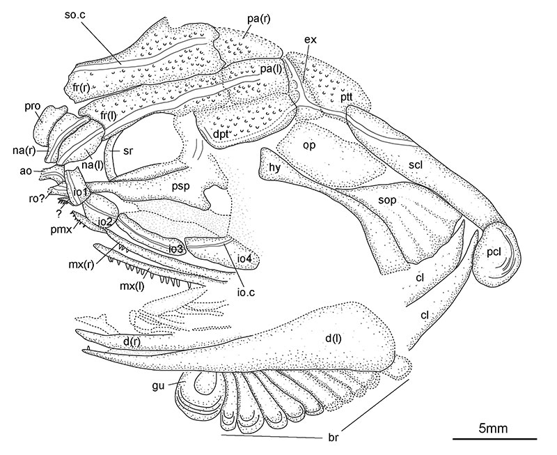
FIGURE 8. Snout bones in †Condorlepis groeberi. 8.1, Line drawing of the slightly disarticulated postrostral, nasal and antorbital bones in MPEF-PV 3958. 8.2 Line drawing of the articulated postrostral and nasal bones in MPEF-PV 1732B.
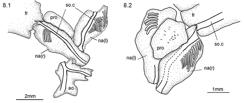
FIGURE 9. Line drawing of the skull of †Condorlepis groeberi in MPEF-PV 3958.
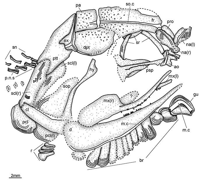
FIGURE 10. Upper jaw of †Condorlepis groeberi. 10.1, Detailed photograph of MPEF-PV 1731B showing the premaxilla; the anterior process of the parasphenoid is also well shown in this picture; scale bar equals 2 mm. 10.2, Disarticulated maxilla in MPEF-PV 1496-5A, scale bar equals 5 mm.
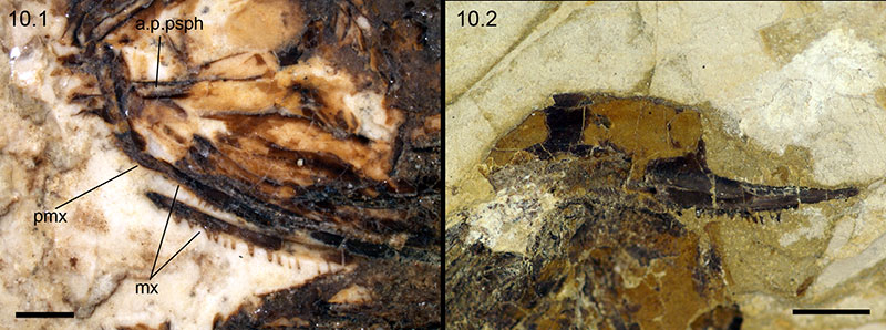
FIGURE 11. Vertebral column of †Condorlepis groeberi. Detailed photographs of the specimen MPEF-PV 1731B: 11.1, anterior portion of the abdominal region, directly behind the skull; 11.2, central portion of the vertebral column showing the transitional morphologies between the abdominal and caudal regions.
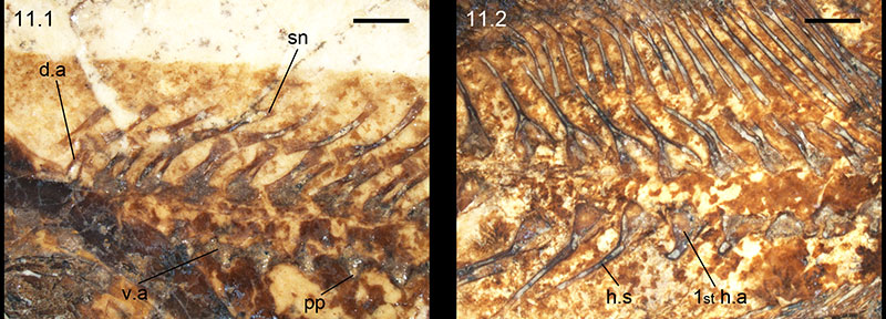
FIGURE 12. Caudal skeleton of †Condorlepis groeberi. 12.1, Photograph of MPEF-PV 1732B; 12.2, photograph of MPEF-PV 3958A. Scale bars equals 2 mm.
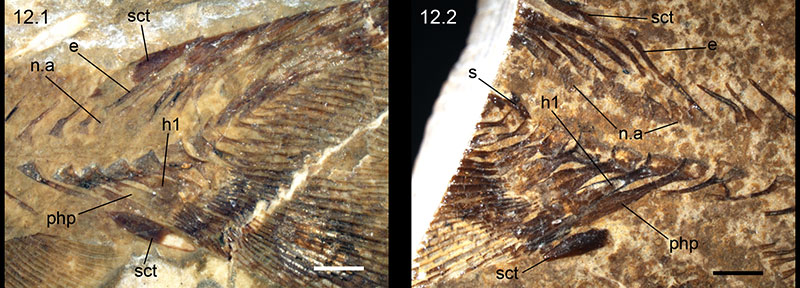
FIGURE 13. Detailed features of the caudal skeleton of †Condorlepis groeberi. 13.1, Photograph of MPEF-PV 1731B showing the exquisitely preserved small cercus-like extension of the body lobe, which is indicated by the black arrow; 13.2, detailed photograph of the ventral margin of the caudal fin in MPEF-PV 3958B showing the distal segments of two marginal fin rays intercalating between the fringing fulcra (black arrows). Scale bars equals 2 mm.
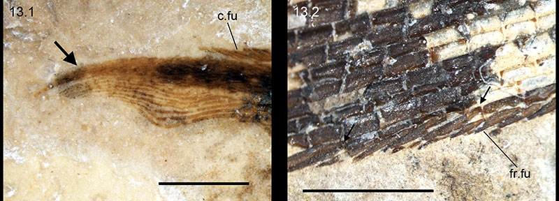
FIGURE 14. Dorsal and anal fins of †Condorlepis groeberi. 14.1, dorsal fin support in MPEF-PV 10506 showing separated proximal and middle radials; 13.2, anal fin support in MPEF-PV 1731B. Scale bars equals 2 mm.
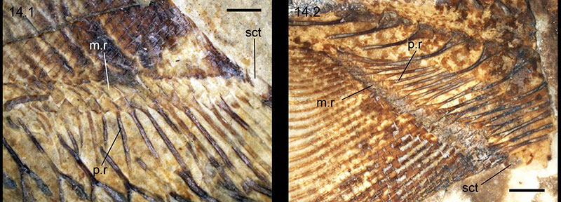
FIGURE 15. Photograph of †Condorlepis groeberi (MPEF-PV 1766) showing the pectoral spine. Scale bar equals 5 mm.
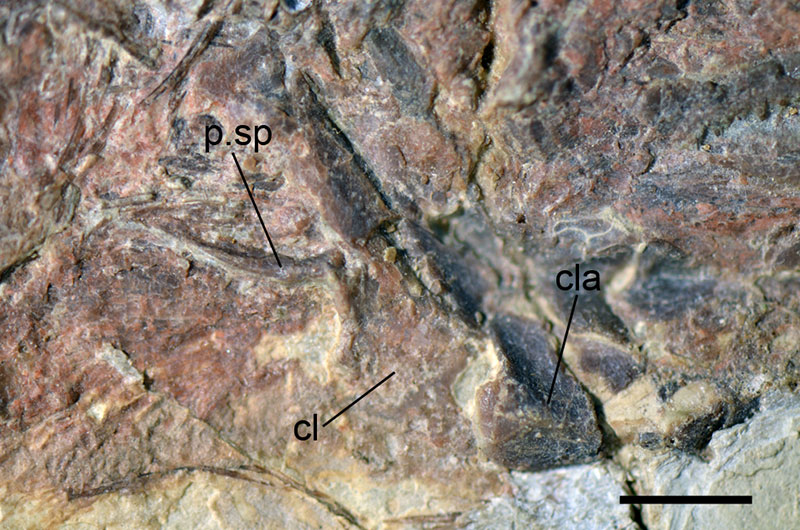
FIGURE 16. Body squamation in †Condorlepis groeberi (MPEF-PV 1556-6). Scale bar equals 5 mm.
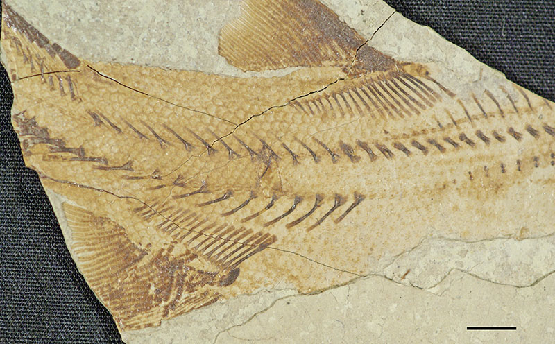
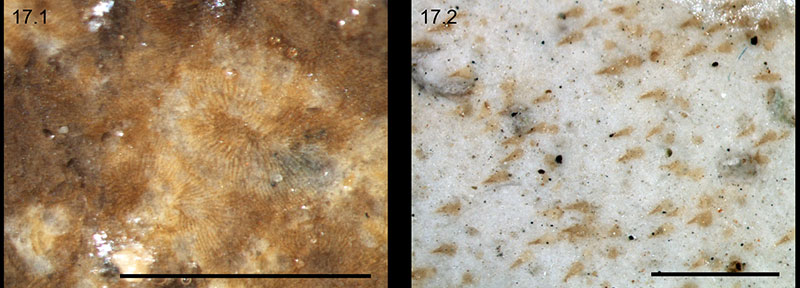
FIGURE 17. Detailed features of the scales in †Condorlepis groeberi. 17.1 Photograph of scales in MPEF-PV 1558 showing the ridges radiating from the growth center towards the borders of the scales; 17.2 photograph of scales in MPEF-PV 1556-6 showing the very fine and pointed denticles irregularly arranged. Scale bars equal 1 mm.