FIGURE 1. (1) Geological sketch of the Iberian Peninsula showing the setting of the Cantabrian Mountains. (2) Geological setting of the study area in Tanes, Cantabrian Mountains. (3) Stratigraphic log of the lower-middle Cambrian transition in the Cantabrian Mountains.
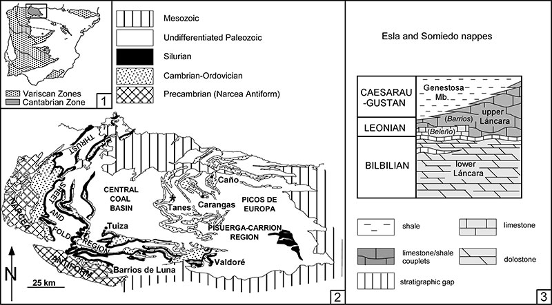
FIGURE 2. Principal parameters measured in dorsal (1) and lateral (2) views of Hadimopanella oezgueli sclerites from the Genestosa Member. Abbreviations: largest / shortest diameter of outline sclerite (Dmax/Dmin), middle surface (d´max/d´min) and tuberculated surface (dmax/dmin); height of marginal brim (h1), middle surface (h2), and tuberculated surface (h3); slope of marginal brim (α1), middle surface (α2), and tuberculated surface (α3); radius base (r).
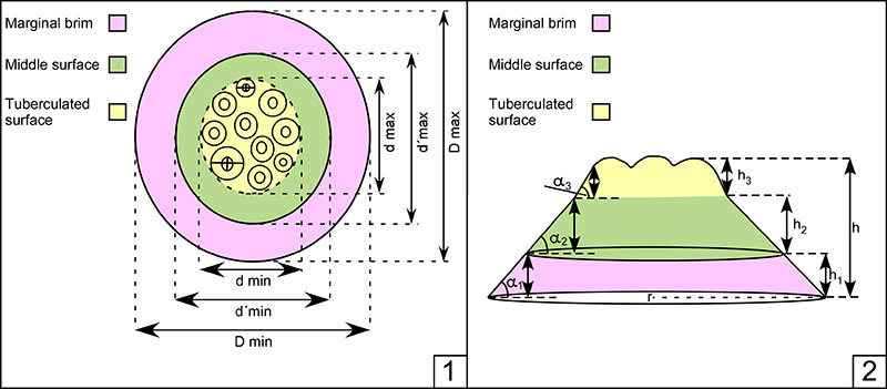
FIGURE 3. (1) Diameter frequency histogram (maximum diameter of outer surface, D max) of dorsal-view sclerites (n=33). (2) Height frequency histogram (from base to the tubercle tip) of lateral-view sclerites (n=36).
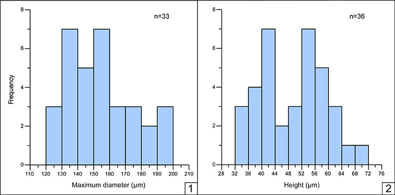
FIGURE 4. (1) Cluster diagram of dorsal-(1) and lateral-view (2) sclerites; morphotypes A1, A2, B1, and B2 are identified in (1), whereas no morphotypes can be recognized in (2).
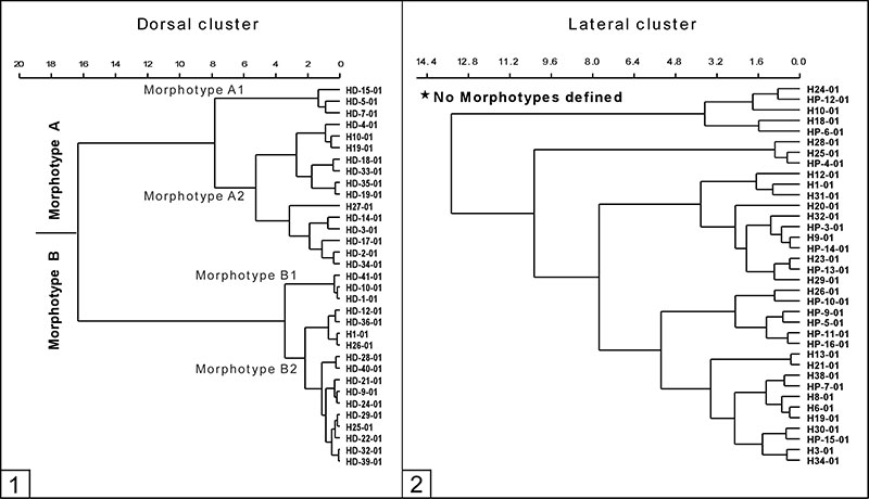
FIGURE 5. (1 to 16) SEM photographs of Hadimopanella oezgueli Gedik, 1977 sclerites from the Genestosa Member, middle Caesaraugustan. (1-2) Morphotype A1; (3, 6-7, 10) Morphotype A2; (11) Morphotype B1; (9, 14-16) Morpthotype. B2. (4-5, 8, 12-13) Lateral-view of sclerites; scale bars for dorsal views equal 50 µm and for lateral views scale bars equal 100 µm (except no. 13 =50µm). Illustrated specimens are housed in the Instituto Geológico y Minero de España (IGME: MGM prefix), Spain: from MGM 1118K to MGM 1133K.
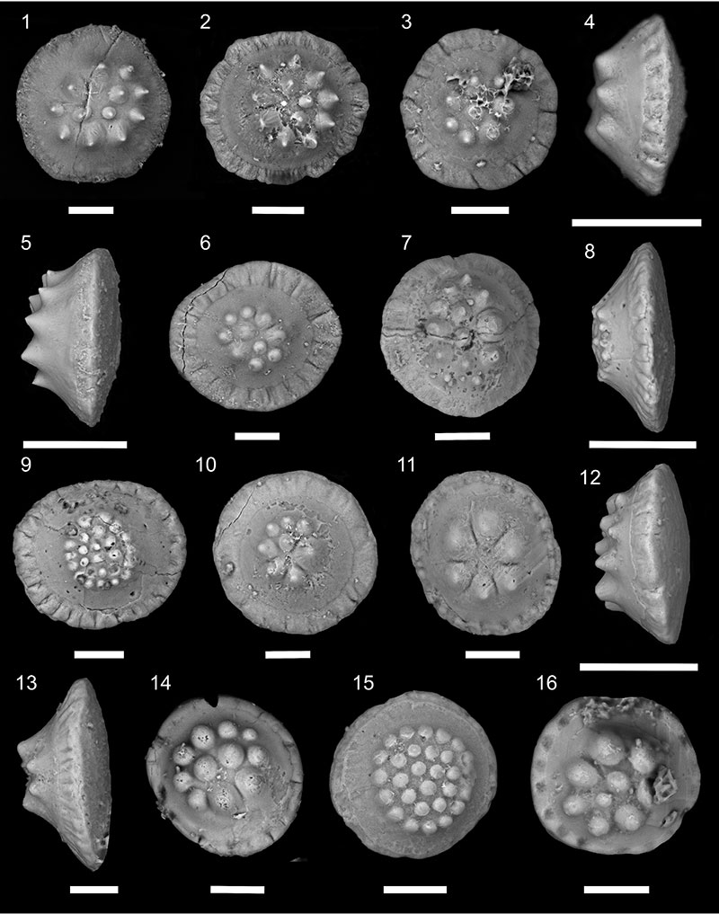
FIGURE 6. (1) Principal-coordinates diagram of dorsal-view sclerites from the Genestosa Member compared with other occurrences of Hadimopanella oezgueli Gedik, 1977 from Gondwana and Siberia. (2) Principal-coordinates diagram of lateral-view sclerites from the Genestosa Member with indication of “ Hadimopanella knappologica ”-type sclerites.
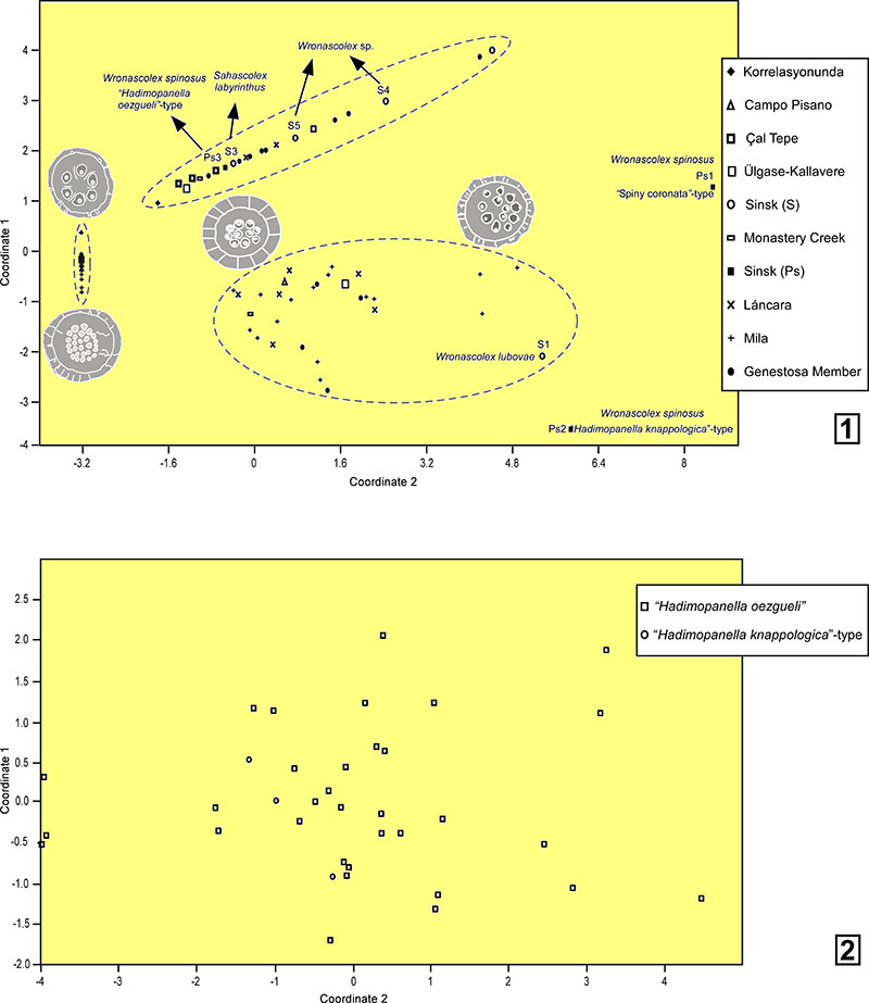
FIGURE 7. (1-3) Dorsal views of sclerites with eroded tubercles; only specimen 2 preserves its original marginal brim. Scale bars equal 50 µm (MGM 1134K to MGM 1136K).
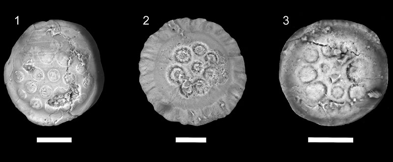
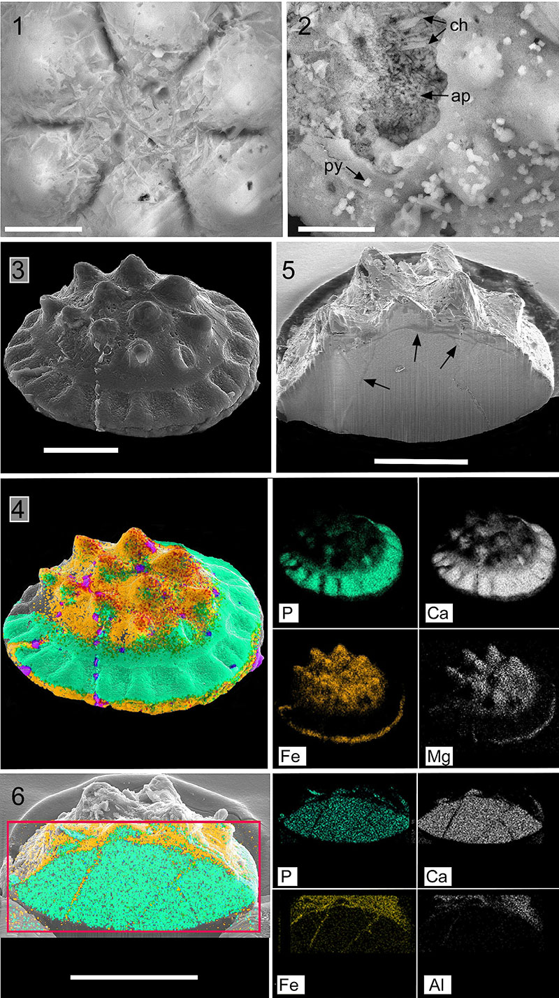
FIGURE 8. Fig. (1) Honey-comb arrangement of chlorites on the top of a sclerite (detail of Figure 5.11) MGM 1128K. (2) Chlorites embedded in the apatite (ap) framework of a corroded sclerite (arrowed) with diagenetic crystals of pyrite (py) and chlorite (ch), MGM 1104K. (3) Complete sclerite, MGM 1137K. (4) BSE analysis of previous sclerite with chlorite arrangement marking porous and fissured areas. (5). Lateral section of sclerite showing fissure network (arrowed), MGM 1138K. (6) BSE analysis of previous sclerite with chlorites occluding the internal fissure network. Scale bars: 1-2 equal 20 µm; 3-4 equal 100 µm; 5 equals 40 µm; 6 equals 60 µm.