FIGURE 1. Sketch of the measurement techniques in micro-CT device.
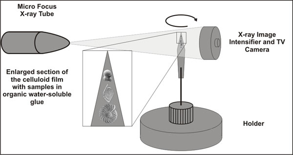
FIGURE 2. Workflow diagram of acquisition and numerical and visual rendering with proposed software packages (modified after SkyScan 2005).
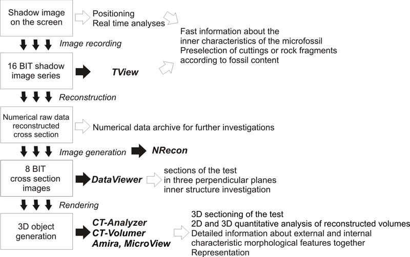
FIGURE 3. Specimens with different wall material fixed with gomme adragante (water-soluble glue) on celluloid films. Bottom to top: Spirillina infima (Strickland, 1846) (Jurassic, Som Hill, Transdanubian Range, Hungary), Paalzowella scalariformis Paalzow, 1917 (Jurassic, Som Hill, Transdanubian Range, Hungary), Bolivina dilatata Reuss, 1850 (Badenian, North Hungarian Range). The largest diameter is 300 μm.
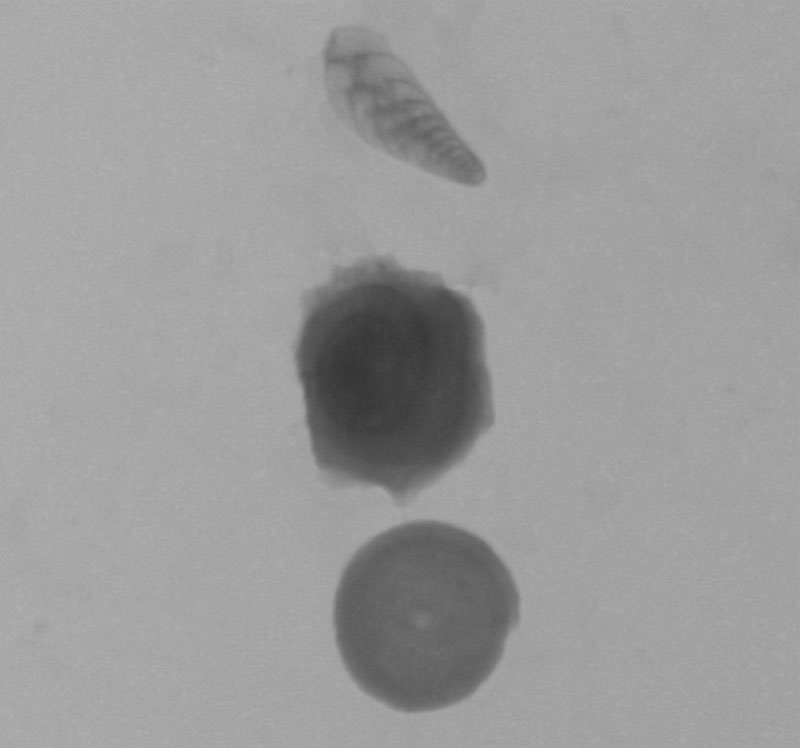
FIGURE 4. X-ray shadow images of (4.1) hyaline Elphidium macellum (von Fichtel and Moll, 1798) (Sarmatian, Zsámbék Basin, North Hungary) (the largest diameter is 400 μm) and (4.2) agglutinated Tritaxia tricarinata (Reuss, 1844) (Cretaceous, Magyarpolány, Transdanubian Range) test (the largest diameter is 250 μm) of foraminifera.
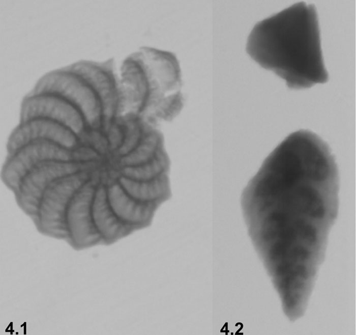
FIGURE 5. X-ray shadow image of the Badenian (Székkutas, SE-Hungary) cuttings in plastic tube holder. Red circles indicate the foraminifera. The largest diameter of the foraminifera is 300 μm.
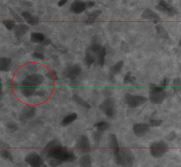
FIGURE 6. 3D model of Triloculina schreiberiana d'Orbigny, 1839 (Recent, Zadar, Adriatic Sea), side view (Amira software). The largest diameter is 350 μm.
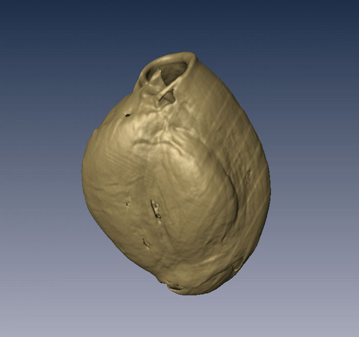
FIGURE 7. 3D model of Triloculina schreiberiana d'Orbigny, 1839 figured on Figure 6, with translucent last chambers, side view. (Amira software). The largest diameter is 350 μm.
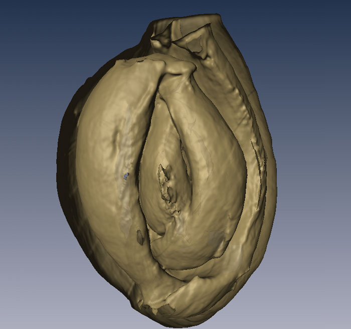
FIGURE 8. Section across the embryonic chamber of the 3D models of Triloculina schreiberiana d'Orbigny, 1839 illustrated on Figure 6 (Amira software). The largest diameter is 350 μm.

FIGURE 9. 3D model of of Triloculina schreiberiana d'Orbigny, 1839 illustrated on Figure 6, apertural view (Amira software). The largest diameter is 350 μm.
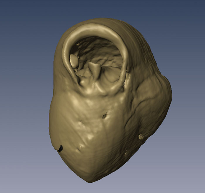
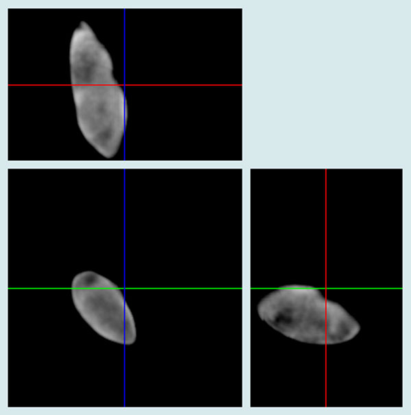
FIGURE 10. Apertural section penetrating the embryonic chamber (large image) of Spiroloculina cymbium d'Orbigny, 1839 (Recent, Zadar, Adriatic Sea) and three perpendicular views (right side) (MicroView software). The largest diameter is 350 μm.
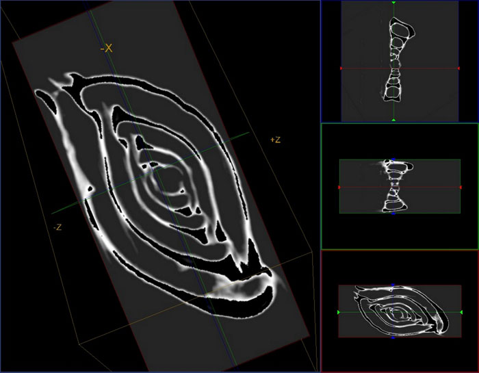
FIGURE 11. Section across the embryonic chamber of the 3D models of Spiroloculina cymbium d'Orbigny, 1839 figured on Figure 10 (Amira software). The largest diameter is 350 μm.
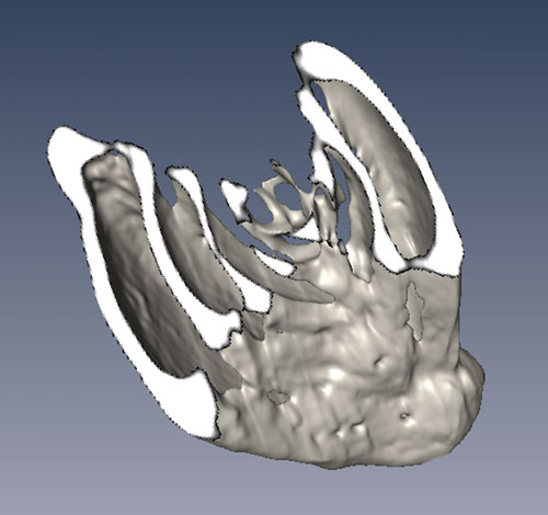
FIGURE 12. 3D model of of Spiroloculina cymbium d'Orbigny, 1839 illustrated on Figure 10, apertural view (Amira software). The largest diameter is 350 μm.
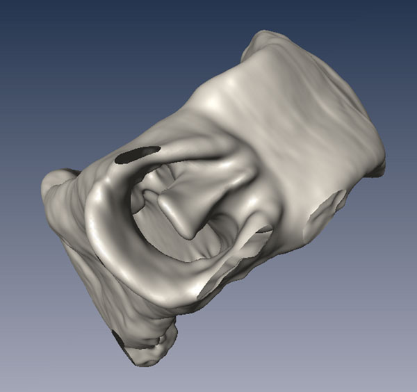
FIGURE 19. Axial section of Globigerinoides trilobus (Reuss, 1850) (Badenian, North Hungarian Range) and three perpendicular views (right side) (MicroView software). The largest diameter is 200 μm.
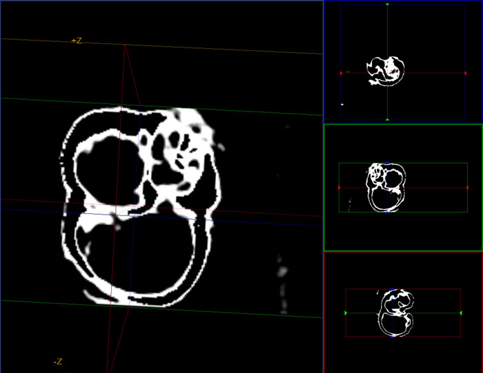
FIGURE 21. Section across the juvenile chambers of the 3D models of Orbulina universa d'Orbigny, 1839 (Badenian, North Hungarian Range) (Amira software). The largest diameter is 150 μm.
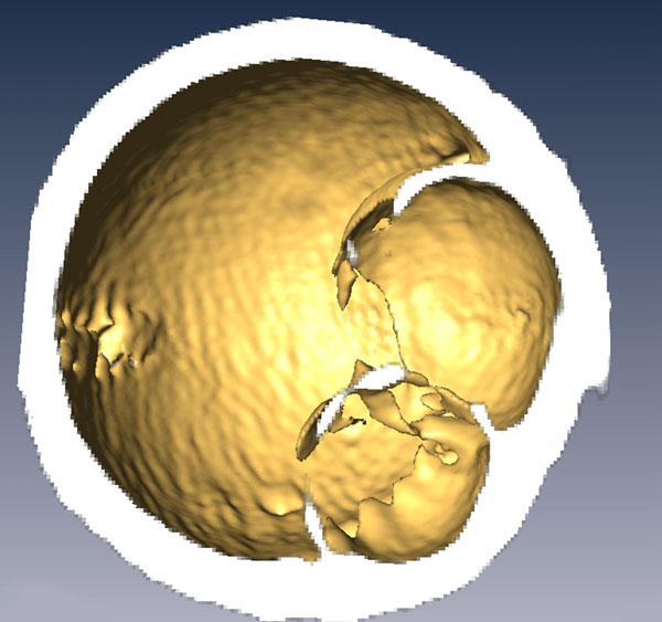
FIGURE 23. Cross section images of Carpathian (North Hungarian Range) (23.1) and Sarmatian (Zsámbék Basin, Hungary) (23.2) washing residue in plastic tube holder (DataViewer software). Diameter of the plastic tube is 3.5 mm.
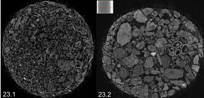
FIGURE 25. Sectioning in three perpendicular planes of recrystallized test and infilled by sparrycalcite of Eoguttulina sp. (Cretaceous, Calvaria Hill, Transdanubian Range) (DataViewer software). The height of the test is 310 μm.