 Figure
1 shows Cambrian chancelloriids from the Burgess Shale and Wheeler Shale.
The specimen from the Burgess Shale (Figure
1A) is preserved both in its soft body and in the bristly exoskeleton
consisting of spiny sclerites. The specimen from the Wheeler Shale (Figure
1B) is preserved as limonitic sclerites, without apparent remaining soft
tissue. Figures 1A1 and 1B1
are taken in plain light. Although the specimens are flattened in the shales,
the images in figures 1A2
and 1B2 appear
three-dimensional because of the photographic techniques used to maximize the
contrast between the fossils and the surrounding matrix. These images have
been produced by a combination of techniques that are generally available, but
remarkably under-used in fossil photography. None of the techniques is new. The
present article is intended to show their usefulness to palaeontologists and
make them more widely known and applied. Parts of the procedures would be
difficult to carry out without digitized images and graphic tools such as those
included in Adobe Photoshop®, but the principles involved predate the invention
of digital imaging.
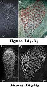
Figure
1 shows Cambrian chancelloriids from the Burgess Shale and Wheeler Shale.
The specimen from the Burgess Shale (Figure
1A) is preserved both in its soft body and in the bristly exoskeleton
consisting of spiny sclerites. The specimen from the Wheeler Shale (Figure
1B) is preserved as limonitic sclerites, without apparent remaining soft
tissue. Figures 1A1 and 1B1
are taken in plain light. Although the specimens are flattened in the shales,
the images in figures 1A2
and 1B2 appear
three-dimensional because of the photographic techniques used to maximize the
contrast between the fossils and the surrounding matrix. These images have
been produced by a combination of techniques that are generally available, but
remarkably under-used in fossil photography. None of the techniques is new. The
present article is intended to show their usefulness to palaeontologists and
make them more widely known and applied. Parts of the procedures would be
difficult to carry out without digitized images and graphic tools such as those
included in Adobe Photoshop®, but the principles involved predate the invention
of digital imaging.
Technical Note. The photographs in this article (except for Figure 9) were taken with a Leaf Microlumina digital camera equipped with a Nikon AF Micro Nikkor 60 mm macrophoto lens and extension bellows. Polarizing filters were applied to the lens and light sources as described later. For the originally captured raster images, the highest available resolution (3,380×2,700 pixels) was used. The images were processed on an Apple Macintosh® computer using Adobe Photoshop (version 5.0). Blending modes of layers and channels were applied as described for each picture; adjustment of input and output levels and application of the unsharp mask filter were performed in all cases to optimize the pictures, but the specific settings of these filters are not given for the individual pictures. (See Basic Concepts for a brief explanation of raster imaging, particularly as it applies to work in Adobe Photoshop.) No retouching was done on any of the images.
Museum number prefixes for the illustrated specimens are as follows: NHM = Natural History Museum (London); ROM = Royal Ontario Museum, Toronto; USNM = National Museum of Natural History, Washington, DC; NIGPAS = Nanjing Institute of Geology and Palaeontology, Academia Sinica; NRM = Swedish Museum of Natural History, Stockholm; PMU = Museum of Palaeontology, Uppsala University; and LO = Department of Historical Geology and Palaeontology, Lund University.
![]()