USING POLARIZED LIGHT
Light reflected from a surface is differently polarized depending on the
angle of incidence, the optical properties of the material, and the topography
of the surface. If polarized light is used for illumination, changes in the
polarization of the returned light can be analyzed using an additional
polarization filter in front of the detecting device. This is the principle of
epi-polarizing microscopes and other similar instruments. The ability of such
devices to separate directly reflected light from backscattered light is used,
for example, in ophthalmology (Fariza
et al. 1989) and dermatology (Philip
et al. 1988; Anderson 1991; Phillips
et al. 1997).
The principle is eminently useful for fossil photography as well. Rayner
(1992) used it to obtain high-contrast images of coalified fossils in dark
shales, and Boyle (1992) applied it
to Burgess Shale fossils. The technique is simple. In the setup used here, the
camera lens is fitted with a regular polarizing filter, and spot lamps used for
the illumination are also provided with polarizing filters that can be rotated
in front of the lamps (filters of the appropriate size can be cut from
commercially available gelatin filters). The filter at each of the light sources
is then rotated individually so as to obtain maximum extinction of reflections
from the object (or a reflecting object temporarily inserted in front of the
camera lens); this is most easily done if the other light sources are covered or
put out when the filter of one source is adjusted. The procedure is analogous to
crossing the nicols in a petrographic microscope and will consequently be
referred to here as crossed nicols (the term nicol in current
usage refers not only to a Nicol prism, but to any filter that polarizes light).
Further practical considerations are discussed by Rayner
(1992) and Boyle (1992).
With this setup, dramatic contrasts may be obtained from otherwise very
low-contrasting material, depending on whether the light at reflection keeps its
original polarization or becomes more or less strongly repolarized. Also,
because direct reflections are repressed, the effect is similar to that obtained
when a specimen is immersed in water or some other clear fluid. Both these
effects are very useful when photographing fossils from two of the classic
Cambrian preservation lagerstätten, the Burgess Shale and the Maotianshan
mudstone (with the Chengjiang fauna), as well as other fossils, such as
graptolites and plants, preserved in shales or mudstones.
Burgess Shale
The Middle Cambrian Burgess Shale in British Columbia is not only famous for
its exquisitely preserved fossils, but also infamous for the difficulties it
presents to the photographer. The fossils are generally preserved in a shiny
film that differs only slightly in colour from the surrounding rock. Commonly
interpreted as an aluminosilicate film (Conway
Morris 1977; Whittington 1985; Conway
Morris 1990; Towe 1996; Orr
et al. 1998), its reflectant matter appears to consist mainly of thermally
altered organic carbon (Butterfield
1996). The reflectance of this film makes it possible to photograph the
fossils by tilting them so that the directly reflected light (ultraviolet light
is commonly used for increased contrast) falls into the camera lens (Conway
Morris 1985). The same property, however, also allows us to make use of
polarized light to increase the contrast between fossils and shale (Boyle
1992).
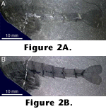 In
Figure 2, a specimen of Waptia
(cf. Briggs et al. 1994, pp. 157–158)
from the Burgess Shale has been immersed in water and photographed without (Figure
2A) and with (Figure 2B) crossed
nicols. The image taken without crossed nicols shows the low contrast between
the dark shale and the films representing the fossil soft parts. When crossed
nicols are applied (Figure 2B), the
improvement is dramatic: the outlines of the soft parts are now clearly visible
against the shale surface.
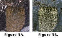 The
same procedures were applied to the images in Figure
3, showing the sponge Vauxia (cf. Rigby
1986) from the Burgess Shale. The details of the organic skeleton are
considerably enhanced under crossed nicols (Figure
3B).
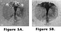
Figure 4 and Figure
5 show grey-scale images of two more Burgess Shale fossils, Marrella
(cf. Whittington 1971) and Burgessia
(cf. Hughes 1975). In Figure 4A and Figure
5A, the specimens have been immersed in water and photographed without
crossed nicols. (The dark irregular patch in Figure
4 is squeezed-out internal fluids and/or decomposed body tissues, a common
occurrence with Marrella.) In Figures 4B
and 5B, the specimens are photographed
with crossed nicols. All four pictures represent a single colour channel.
 A
number of high-quality photographs of Burgess Shale fossils have been produced
throughout the years; see, for example, the photograph of Thaumaptilon by
B.K. Harvey in Conway Morris (1993),
figures 1–2, the suite of photographs by C. Clark in Briggs
et al. (1994), the ctenophore images by several photographers (including B.
Boyle) in Conway Morris and Collins
(1996), or the figures of Alalcomenaeus in Briggs
and Collins (1999). These have been taken using various methods, including
ultraviolet radiation, direct reflections, low-angle lighting, water immersion,
and crossed nicols.
Chengjiang
The early Cambrian Maotianshan mudstone in south China, containing the
exquisitely preserved Chengjiang fauna (e.g., Hou
and Bergström 1997), represents a different problem for photography than
the Burgess Shale. Although flattened, the fossils are preserved in considerably
higher relief than those of the Burgess Shale. Consequently, low-angle light can
bring out good details. Also, there is commonly a colour difference between
fossils and matrix, brought out by iron-rich red films representing part of the
soft bodies. The main problem in photographing them is, instead, that the
mudstone is very friable and cannot be immersed in a fluid without breaking
apart. Thus the common technique of photographing fossils immersed in water,
glycerin or some other suitable liquid to remove reflections and increase
contrast cannot be used. This is where the technique of polarizing the light
comes in useful. (The red colour of the Chengjiang fossils also makes them
suitable for photography with orthochromatic films, which are insensitive to
red, as shown in the photographs by U. Samuelsson in Hou
and Bergström 1997.)
 Figure
6 shows a specimen of Yunnanozoon from the Chengjiang mudstone (cf. Hou
et al. 1991; Chen et al. 1995; Dzik
1995; Shu et al. 1996). Figure
6A is taken without, and Figure 6B
with, crossed nicols. The difference in result is less dramatic than in the case
of the Burgess Shale fossils; however, the use of crossed nicols has an effect
similar to that of immersing the specimen in liquid, namely to reduce
reflections and enhance contrasts.
Other carbonized fossils
The effects of using polarized light for the photography thus will range from
good to spectacular, depending on the differences in reflectance of the objects.
Only experimentation will tell how useful the method is in any particular case,
but carbonized fossils seem consistently to yield fine results, as noted by Rayner
(1992).
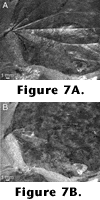
 Two
further examples of such fossils are given here. Figure
7 shows a Tertiary leaf from Spitsbergen. In this case, the surface
topography of the leaf comes out best in unpolarized light (Figure
7A), whereas the use of crossed nicols brings out the contrast with the
matrix as well as the colour differences within the leaf (Figure
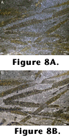
7B). Figure 8 shows graptolites
from Ordovician grey shales of Scania, Sweden. Although there is a colour
difference between fossils and matrix that comes out without crossed nicols (Figure
8A), a clear contrast is not obtained until crossed nicols are applied (Figure
8B).

 In
Figure 2, a specimen of Waptia
(cf. Briggs et al. 1994, pp. 157–158)
from the Burgess Shale has been immersed in water and photographed without (Figure
2A) and with (Figure 2B) crossed
nicols. The image taken without crossed nicols shows the low contrast between
the dark shale and the films representing the fossil soft parts. When crossed
nicols are applied (Figure 2B), the
improvement is dramatic: the outlines of the soft parts are now clearly visible
against the shale surface.
 The
same procedures were applied to the images in
  In
In  A
number of high-quality photographs of Burgess Shale fossils have been produced
throughout the years; see, for example, the photograph of Thaumaptilon by
B.K. Harvey in

 Two
further examples of such fossils are given here.