![]()
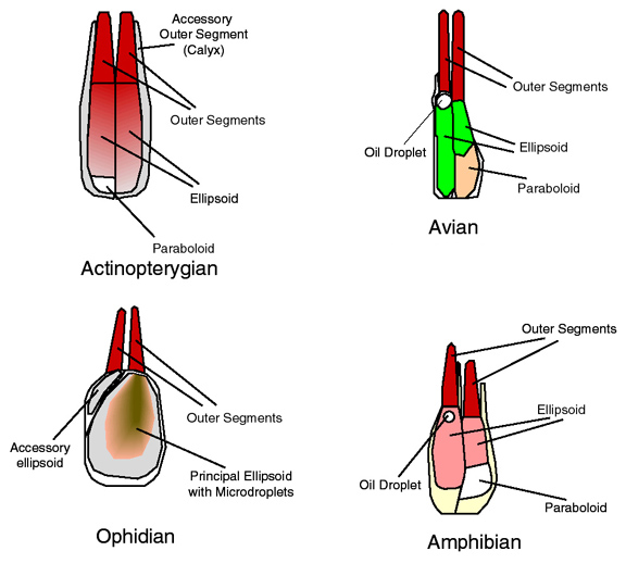
Figure 11. Double cone morphologies. These schematized drawings depict some of the variety in constitution of double cones. Diagrams only show inner and outer segments of photoreceptors (the cells also contain myoids and nuclear regions). Inner segment organelles generally provide metabolic and/or optical functions. For instance, the ellipsoids are comprised of dense aggregations of mitochondria, which not only serve as sites for the conversion of sugars to other fuels useful to the cells, but also have a high refractive index and thus tend to guide light into the outer segments. One feature commonly shared among double cones is the direct apposition of the ellipsoids against the membranes which unite the two constituent photoreceptors of a given double cone. This suggests that the doubling of photoreceptors arose as a means to optically couple the joined photoreceptors. Exactly what benefit such coupling might serve is presently unknown. Microdroplets in snake cones may be descended from oil droplets more similar to those currently found in other reptilian retinas. (Avian cones derived from images in Walls 1942, and Morris and Shorey 1967; amphibian derived from Nilsson 1964; ophidian after Walls 1942, and Wong 1989).