![]()
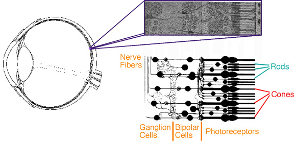
Figure 3. The vertebrate retina. The retina is the thin set of tissue layers lining the inside of the vertebrate eye. The drawing to the left is a cross-section of a human eye. The drawings to the right indicate the position and orientation of the three layers of cell bodies in the neural portion of the retina. The last layer -- sometimes referred to as the bacillary layer or the photoreceptor layer -- contains the cells which convert the absorption of light into an electrochemical signal. Almost all vertebrate retinas contain two major populations of photoreceptors, rods and cones. Although originally so named because of their shapes in many animals, modern usage generally discriminates between the two populations on functional grounds. Rods are active at low light levels, and are thus saturated and useless during the day. Conversely, cones are less sensitive, and thus operate almost exclusively during the day (or for modern humans under artificial sources of illumination). Cones can usually be further subdivided into a small number of classes based upon their spectral sensitivity. (Drawings adapted from Walls 1942)