![]()
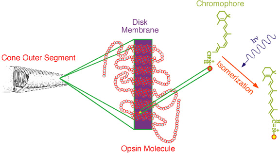
Figure 5. Phototransduction. The drawing to the left is reproduced from Figure 4. The diagram in the center schematically depicts how a photopigment is embedded within a disk membrane of a photoreceptor outer segment. Each circle represents an individual amino acid. The filled yellow circle is a lysine to which is attached the chromophore, the molecule schematically represented on the right. The chromophore is a vitamin A derivative which can exist in two stable configurations. The absorption of a photon may cause the chromophore to convert from the 11-cis form to the all trans form as shown by the chemical reaction. This conformational change in the chromophore causes a change in the shape of the opsin molecule. The opsin's change in shape converts it from an inactive to an activated enzyme, and thus is light absorption converted to a biochemical signal in a photoreceptor. (Opsin sequence adapted from Wilkie et al. 1988).