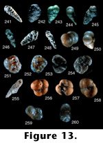
 FIGURE
13.241-260.
FIGURE
13.241-260.Textularia intosiana Nakamura (continued from 12.240).
13.241. Edge view showing aperture, 7.5x (LD = 3.06 millimeters).
Textularia notoensis Asano (1953a, p. 20, pl. 1, figures 4 and 5).
13.242. Side view showing aperture, 10x (LD = 2.60 millimeters).
Photograph of holotype (#75268) in the Tohoku University collections. This is a very coarse-grained species that looks like a distinct species.
Textularia sintikuensis Nakamura (1937, p. 135, pl. 10, figure 7a and 7b).
13.243. Side view, 45x (LD = .67 millimeters).
13.244. Edge view showing aperture, 45x (LD = .67 millimeters).
Photographs of holotype (#60858) in the Tohoku University collections. This is a distinct species.
Textularia taiwanica Nakamura (1937, p. 135, pl. 10, figure 8a and 8b).
13.245. Slanted side view, 13.5x (LD = 2.00 millimeters).
13.246. Edge view showing aperture, 13.5x (LD = 2.00 millimeters).
Photographs of holotype (#60859) in the Tohoku University collections. This species was placed in synonymy with T. foliacea Heron-Allen and Earland by Huang (1968).
Textularia uedai Asano (1936e, p. 611, pl. 30, figure 1a and 1b).
13.247. Side view, 13.5x (LD = 2.07 millimeters).
13.248. Edge view showing aperture, 13.5x (LD = 2.07 millimeters).
Photographs of holotype (#21363) in the Tohoku University collections. This species was placed in synonymy with T. foliacea by Asano (1950c).
Tosaia hanzawai Takayanagi (1953, p. 30, pl. 4, figure 7a and 7b).
13.249. "Dorsal" view, 45x (LD = .47 millimeters).
13.250. Apertural view, 45x (LD = .58 millimeters).
Photographs of holotype (#67147) in the Tohoku University collections. This is a prominent species in many deep-sea environments; it is often confused with Eggerella bradyi (Cushman 1911). However, this is a calcareous species, not agglutinated, and so it is distinct. 13.249 is the first picture in which the first five chambers are visible if the photo is enlarged; Takayanagi, the author of this species, had never been able to see the first few chambers of this species until we projected this slide on a screen where it was still in focus even at room size. The value of the SLM was apparent with this species.
Trochammina bullata Takayanagi (1960, p. 85, pl. 4, figure 1a-1c).
13.251. Ventral view, 13.5x (LD = .96 millimeters).
13.252. Dorsal view, 13.5x (LD = .96 millimeters).
Photographs of holotype (#74862) in the Tohoku University collections. Specimens observed in the Holocene of the North Atlantic (Cole 1981; Scott 1987) were referred to this species on the basis of type illustrations. Examination of type specimens established that they were not the same as those observed in North Atlantic sediments, demonstrating the importance of looking at original types.
Trochammina nipponica Asano (1953b, p. 54, figure 5a-5c).
13.253. Ventral side, 13.5x (LD = 1.48 millimeters).
13.254. Dorsal side, 13.5x (LD = 1.48 millimeters).
13.255. Edge view, 13.5x (LD = 1.26 millimeters).
Photographs of holotype (#75288) in the Tohoku University collections. The closest species to this one is T. macrescens Brady (1870). But unlike any other trochamminids, this one has almost a "Cibicides" like appearance because of deformation caused by lateral compression in the rock.
Trochammina nobensis Asano (1951d, p. 8, figures 3 and 4).
13.256. Ventral view, 45x (LD = .49 millimeters).
13.257. Dorsal view, 45x (LD = .49 millimeters).
13.258. Edge view, 45x (LD = .49 millimeters).
Photographs of holotype (#67101) in the Tohoku University collections. This species was subsequently determined to be a planktic foraminifera, Neogloboquadrina nobensis, by Takayanagi and Hasegawa (1987). It is illustrated here to avoid any confusion that might exist.
Trochammina yubarensis Takayanagi (1960, p. 87, pl. 4, figure 4a-4c).
13.259. Ventral view, 45x (LD = .44 millimeters).
Photographs of holotype (#74865) in the Tohoku University collections. This is not a Neogene form, but because it was described in the same publication as T. bullata, it is included here. It is certainly not like any Neogene form.
13.260. Dorsal view, 45x (LD = .44 millimeters).
Note: Jung (1988) has made a comprehensive study of all Japanese forms of the uvigerinids using a variety of characters to describe them, including toothplates, wall structure, and internal structures. He obviously looked at many more specimens than in this study so our opinions are based on some of his observations. However, Scott still feels that some of these forms are close to U. Peregrina, a form never mentioned by Jung.
Uvigerina akitaensis Asano (1950b, p. 14, figures 60-62).
14.261. Side view, 13.5x (LD = 1.33 millimeters).
Photograph of holotype (no number) in the Tohoku University collections. Scott places this species into U. peregrina Cushman (1923). However, we did not examine internal structures as did Jung (1988), who placed this species in the genus Euuvigerina on the basis of toothplate characteristics.
Uvigerina asanoi Matsunaga (1963, p. 113, pl. 42, figure 10a and 10b).
14.262. Side view, 13.5x (LD = 1.33 millimeters).
Photograph of holotype (#85260) in the Tohoku University collections. This species was placed in synonymy with U. akitaensis by JJung (1988).
Uvigerina peregrina Cushman shiwoensis Asano (1958, p. 35, pl. 6, figures 5-8).
14.263. Side view, 13.5x (LD = .96 millimeters).
Photograph of holotype (#77174) in the Tohoku University collections. Jung (1988) elevated this to species status (Euuvigerina shiwoensis), suggesting that this species was much closer to E. aculeata (dOrbigny 1846) than to U. peregrina,S but nevertheless a distinct species.
Uvigerina pseudoampullacea Asano (1938c, p. 613, pl. 17(6), figures 28 and 29).
14.264. Slanted side view, 13.5x (LD = .89 millimeters).
Photograph of holotype (#21428) in the Tohoku University collections. Jung (1988) commented that this species is similar to many spinose forms, but is distinct. Scott agrees that it is similar to many described spinose forms, but cannot say if it is a junior synonym of any older species. Jung (1988) placed this species into Neouvigerina.
Uvigerina schencki Asano (1950b, p. 17, figures 74 and 75).
14.265. Side view, 13.5x (LD = 1.04 millimeters).
Photograph of holotype (#66939) in the Tohoku University collections. Jung (1988) placed this species into the genus Euuvigerina.
Uvigerina substriata Asano (1938c, p. 614, pl. 17(6), figures 21 and 22).
14.266. side view, 13.5x (LD = 1.48 millimeters).
Photograph of holotype (#21430) in the Tohoku University collections. This species was not discussed by Jung (1988), but it appears to be distinct from the other Japanese uvigerinids.
Uvigerina urnula d'Orbigny shiiyaensis Matsunaga (1963, p. 114, pl. 43, figure 4a and 4b).
14.267. Side view, 13.5x (LD = 1.11 millimeters).
Photograph of holotype (#85266) in the Tohoku University collections. This may be a distinct subspecies.
Uvigerina yabei Asano (1938c, p. 613, pl. 17(6), figures 1 and 2).
14.268. Edge view, 13.5x (LD = 1.78 millimeters).
Photograph of holotype (#21429) in the Tohoku University collections. Jung (1988) separated this species from U. akitaensis on the basis of costae and larger size of this species but, earlier in the same paper, admitted that these may not be valid criteria. Jung pointed out that there has been much confusion in Japan over this species, and it is not hard to see why.
Uvigerinella quadrata Iwasa (1955, p. 17, text figure 3a-3c).
14.269. Apertural view, 45x (LD = .42 millimeters).
14.270. Side view, 45x (LD = .47 millimeters).
Photographs of holotype (#65503) in the Tohoku University collections. This looks like a distinct species.
Valvulinera masudai Asano (1953a, p. 20, figure 16a-16c).
14.271. Ventral view, 13.5x (LD = 1.11 millimeters).
14.272. dorsal view, 13.5x (LD = 1.11 millimeters).
Photographs of holotype (# 75270) in the Tohoku University collections. It is probably a distinct species, but it is a badly etched specimen, making it difficult to evaluate.
Valvulinera sadonica Asano (1951a, p. 8, figures 55-57).
14.273. Slanted ventral side, 13.5x (LD = 1.18 millimeters).
14.274. Dorsal view, 13.5x (LD = 1.26 millimeters).
14.275. Edge apertural view, 13.5x (LD = 1.26 millimeters).
Photographs of paratype (#67179) in the Tohoku University collections. This is a very interesting species that has some rare features for this genus, such as the slightly covered umbilical area and what appears to be almost a tubular extension into the umbilical area.
Virgulina akitaensis Iwasa (1955, p. 17, text figure 2a and 2b).
14.276. Apertural side view, 45x (LD = .42 millimeters).
Photograph of holotype (#65502) in the Tohoku University collections. This species is very close to Fursenkoina fusiformis (Williamson 1858).
Virgulina complanata Egger fugeshiensis Asano (1953a, p. 20, pl. 2, figure 22).
14.277. Side view showing aperture, 13.5x (LD = 1.85 millimeters).
Photograph of holotype (#75269) in the Tohoku University collections. The slide containing this specimen was labeled as V. notoensis, but the species name was changed to fugeshiensis in the publication. This species is listed by Takayanagi and Hasegawa (1987), as belonging to the genus Fursenkoina; however, it does not appear to be a variation of F. complanata, but rather a distinct species.
Virgulina ishikiensis Asano (1949, p. 428, fig.1, nos. 45, 46, 48, and 56).
14.278. Side view showing aperture, 45x (LD = .67 millimeters).
Photograph of holotype (#67041) in the Tohoku University collections. This is another variation of F. fusiformis.
NOTE: LD (longest dimension).
![]()
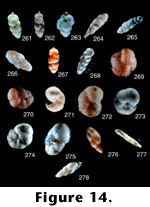 FIGURE
14.261-278.
FIGURE
14.261-278.