RESULTS
Middle Cambrian fossils from the Burgess Shale
have traditionally been photographed in UV light (Conway
Morris 1985). The use of polarizing filters to dampen directly reflected
light has proven to be even more effective in bringing out details not otherwise
visible (Boyle 1992; Bengtson
2000). The application of the reflectance transformation method on a Marella
specimen, preserved mainly as a dark film on dark shale, was highly successful (Fig.
2). The image brightness drops to almost zero when using the low-angle
lights, testifying to the very low relief of fossil features. The method of
specular enhancement leads to a dramatic enhancement of relief, showing detailed
anatomical and taphonomic structure in three dimensions (Fig.
2.3). However, the reduction of color contrast obscures the structures
preserved as dark surface films. The best results were therefore obtained by
combining (adding) the image obtained from specular enhancement with an inverted
version of an unprocessed image made using low-angled virtual light.
Lower Cambrian fossils from the Chengjiang deposit
in the Maotianshan Shale of China (Chen
et al. 1997), preserved with low relief on a light-colored shale, might be
expected to be a good target for PTM imaging. Figure
3 shows a specimen of the sponge Paraleptomitella dictyodroma with
different enhancement techniques. Whereas oblique lighting enhances low-relief
spicules, specular enhancement does not seem to offer any improvement in this
case.
 Low-relief
fern impressions in claystone from the Carboniferous Coal Measures of the Forest
of Dean, Gloucestershire, England, are devoid of color contrast, and can be
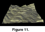
difficult to photograph and study with conventional techniques. Figure
4 shows some results from applying PTM techniques. The 3D reconstruction
from surface normals (Fig. 11) does not
add significant information.
Late Neoproterozoic Ediacaran fossils are commonly
preserved in low relief on the soles or tops of sandstone beds. Imaging them
usually employs whitening in combination with very low-angle lighting. The two
examples illustrated here (Fig. 9, Fig.
10) from the Rawnsley Quartzite of South Australia show that the physically
impossibly low position of the virtual light source through extrapolation of PTM
space may help to bring out faint structures (Fig.
9.3; Fig. 10.3, Fig.
10.4); also that specular enhancement may bring out details that are
otherwise obscured by the granularity of the sandstone (Fig.
9.5–9.9).
PTM imaging of a slab of Cambrian shelly fragments
(hyolithids and trilobites) in a dark shale of the Alum Shale Formation of
Krekling, Norway, illustrates the use of specular enhancement for amplifying
relief and substituting for physical whitening of the specimen (Fig.
5.3).
Fossils of high relief are generally easier to
photograph than those of low relief. Here, traditional photographic methods
usually produce good results using a proper choice of lighting in combination
with surface treatment. The Cambrian trilobite in Fig.
8, from the Alum Shale Formation of the Närke, Sweden, is a classic object
for palaeontological photography. The different orientations of the virtual
light in Fig. 8.2 and Fig.
8.3 serve to illustrate the importance of light directions for the
understanding of the morphology; the full effect is best seen in the interactive
models. As with the specimen in Fig. 5,
specular enhancement may substitute for physical whitening in producing a more
even surface color (Fig. 8.4).
A promising application of PTM imaging is in trace
fossils, where the structures are often faint and oriented in many different
directions so that a single lighting mode cannot capture the full morphology. Figure
6 and Figure 7 show two slabs of
Lower Cambrian trace fossils in "neutral" virtual lighting (1), and
with two directions of the virtual light to demonstrate how differently oriented
structures are picked up in other ways (2, 3). Interestingly, specular
enhancement serves to outline all structures independently of their orientation
because of the strong differences in tone between surfaces nearly parallel to
the main plane and those nearly normal to it (4).
These experiments indicate that reflectance
transformation techniques offer significant improvement over non-transformed
images in some cases, whereas in others the improvement, if any, is marginal
compared with traditional techniques. However, in all cases the possibility of
interactive manipulation of virtual light in PTM images adds a new dimension to
the electronic illustration of fossil specimens.

 Low-relief
fern impressions in claystone from the Carboniferous Coal Measures of the Forest
of Dean, Gloucestershire, England, are devoid of color contrast, and can be
difficult to photograph and study with conventional techniques. Figure
4 shows some results from applying PTM techniques. The 3D reconstruction
from surface normals (Fig. 11) does not
add significant information.