![]()
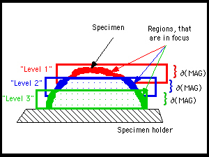
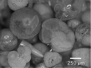
Figure 6. Fusion method. 6.1. Diagrammatic representation of three focal volumes, that intersect a specimen at levels 1 to 3. The height d of a particular focal volume represents the depth of focus and is a function of the magnification. 6.2-6.4. Examples of images taken at three subsequent focal planes. Note various regions with changing sharpness as one focuses from top to down. The images were taken in the monoscopic mode position (=left light beam of the microscope). 6.5. Resulting 'fused' image with extended focus after application of the FocusExtend macro. The images show shells of Recent planktonic foraminifera from surface sediment sample Ki 04, 0-1.5 cm, (Western Mediterranean Sea, lat. 37° 30' N, long. 7° 21' E, water depth 2756 m, taken during French oceanographic expedition VICOMED I in 1986, see Knappertsbusch (1993) for reference). Figures 6 through 9 were constructed from this sample. Click into Figures 6.2-6.5 to obtain enlarged images.
| 6.1. 'Focal volumes' seen in Figs. 6.2-6.4. | ||||
 |
||||
| 6.2. Upper focal plane (Level 1, left). | 6.3. Middle focal plane (Level 2, left). | |||
 |
 |
|||
| 6.4. Lower focal plane (Level 3, left). | 6.5. Fused left image with extended focus. | |||
 |
 |