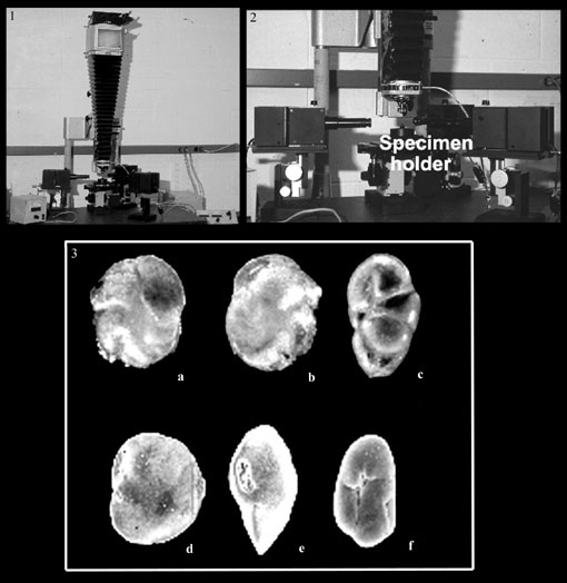
Figure 1. 1.1. Whole view of the Scanning Light Microscope set-up. 1.2. Close view of the SLM set-up. 1.3.a-f. Comparison of Scanning Electron Microscope (SEM) and Scanning Light Microscope (SLM) photographs. a, b: Islandiella teretis (SLM); c: Robertinoides charlottensis (SLM); d, e: Islandiella teretis (SEM); f: Robertinoides charlottensis (SEM) (after Scott and Vilks 1991).