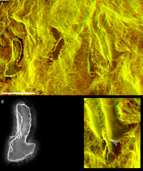
Figure 3. Epoxy replica of the apparatus preserved in conodont specimen RMS GY 1992.41.2 (Royal Museum of Scotland). A Red-green anaglyph stereo image of whole apparatus after acid preparation of the P1 element (at left). B. Close-up of the epoxy replica of the P1 element showing incremental growth lamellae of the crown tissue exposed in the basal cavity (arrowed). C. Red-green anaglyph stereo image close-up of the S3 and S4 elements, showing incremental growth lamellae of the crown tissue exposed along the lower margin of the S4 element (arrowed). Scale bar 0.5 mm.