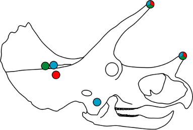
Figure 4. Schematic of Triceratops prorsus skull, showing areas where cranial pathologies are predicted by the SHC (red dots), FHL (blue dots), and OHL (green dots) models of horn locking.