DESCRIPTION
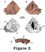 The specimen, MNA V9953, comprises
the major portion of the skull minus the mandible and preserves most of the
marginal tooth rows (Figure 3). However, the surface bone is largely eroded
away or poorly preserved and as a consequence no sutures can be detected. The
skull roof was partially weathered prior to collection of the specimen.
Although the bone is worn and poorly preserved, the general shape of the skull
and the dentition can be discerned, providing the opportunity to make direct
comparisons with known Late Triassic procolophonids.
The specimen, MNA V9953, comprises
the major portion of the skull minus the mandible and preserves most of the
marginal tooth rows (Figure 3). However, the surface bone is largely eroded
away or poorly preserved and as a consequence no sutures can be detected. The
skull roof was partially weathered prior to collection of the specimen.
Although the bone is worn and poorly preserved, the general shape of the skull
and the dentition can be discerned, providing the opportunity to make direct
comparisons with known Late Triassic procolophonids.
As preserved it is a very low flat
skull (Figure 3.3). This might be partially a result of distortion, although
other Late Triassic procolophonids, such as Hypsognathus are also known
to have dorsoventrally compressed skulls.
In dorsal view the skull is
approximately triangular in shape (Figure 3.1). The prominent oval orbitotemporal openings are directed dorsally. These openings are greatly
extended posteriorly, markedly reducing the distance between the posterior
margin of this opening and the posterior margin of the skull roof. Indeed, it
would appear that this distance is approximately equal to the narrowest point
across the frontals. Although the posterior corner of the skull is missing on
the right side, it is possible to partially reconstruct it as the mirror image
of the left side (Figure 3.1). On this basis it is clear that the skull was
broader than it was long. The frontals are constricted slightly toward the
anterior margin of the orbits. A perfectly circular slightly raised area of
matrix occurs along the midline of the skull roof between the anterior
parietals. This area most likely represents the pineal foramen. The posterior
margin of the skull has been eroded, and the braincase is missing.
On the left side the quadratojugal
bears at least three prominent spines (Figure 3.1, 3.3), although additional
spines may have broken off. The jugal extends down below the level of the
maxillary tooth row and its ventral margin appears to slope posteroventrally
(Figure 3.2, 3.3).
The snout is damaged, and it is
difficult to distinguish between the premaxillary and maxillary dentition. The
tooth row is inset from the lateral margin of the skull. Post-mortem distortion
has pushed the tooth row of the left side anteriorly, making it appear that the
right side has more teeth. There appear to be two premaxillary teeth (Figure
3.2), which although damaged, are clearly labio-lingually expanded with a
simple ridged occlusal surface. Four maxillary teeth are preserved on either
side (Figure
3.2). Although the more derived procolophonids tend to exhibit
reduction in marginal tooth numbers, tooth count is not necessarily significant
for phylogenetic analysis. Differences in tooth count are known to represent
ontogenetic variation in other procolophonids (Gow 1977;
Sues and Baird 1998).
The maxillary teeth are
transversely broadened (Figure
3.2). Wear facets on each tooth have obscured
some of the structural details. They appear to possess a labial and a lingual
cusp connected by anterior and posterior transverse ridges that form the margin
of a deep occlusal basin. The anterior margin of the basin is always lower than
the posterior margin. The labial cusp always appears higher than the lingual
cusp, although both of these features could be a result of tooth wear. The wear
patterns are very similar to that seen in an un-named procolophonid from the
fissure deposits at Cromhall Quarry, England (Fraser 1986). However, the
maxillary teeth in the Cromhall form are not as transversely broadened.
Moreover the occlusal basins are considerably deeper in the Abajo form and
comparable to that of Hypsognathus. The Late Triassic Brazilian form Soturnia
also possesses an occlusal depression, although this is manifested as a
prominent anterior-posterior groove on the occlusal surface, not a basin per se
(Cisneros and Schultz 2003). Scoloparia differs greatly in having
maxillary teeth with several cuspules on a single transverse ridge with no
trace of an occlusal basin (Sues and Baird 1998). In palatal view, a bone
extending anterior from the midline probably represents the right vomer (Figure
3.2). It does not preserve any teeth, although this is equivocal, because it is
so poorly preserved. Posterior to the tooth row but anterior of the quadrate
are a thin bone on either side extending posteriorly and slightly towards the
midline (Figure
3.2). They possibly represent the pterygoids, but are too
poorly preserved to confirm this identification.
