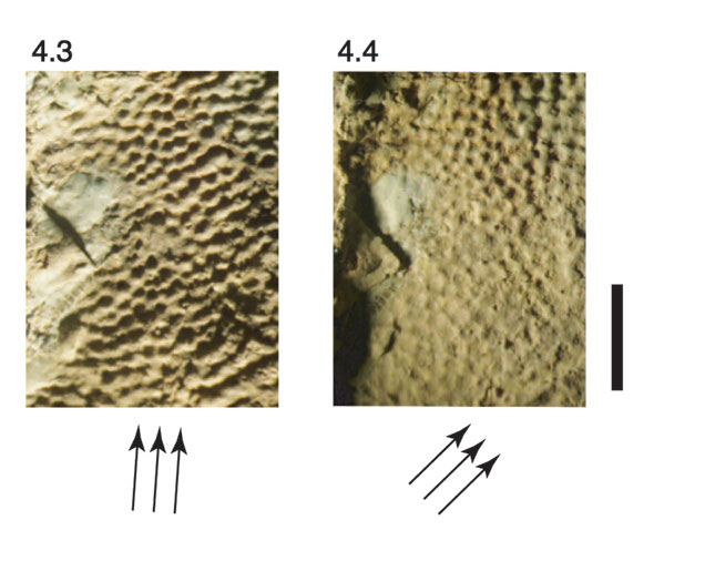
Figure 4. Four micrographs of skin impressions in a shallow theropod track under different laboratory lighting. Arrows depict direction of fiber-optic illumination. Note the uneven illumination, particularly in Figure 4.4. Dimple patterns within a single region appear to vary significantly depending on the positioning of the lamp. In some images, particularly Figure 4.4, these dimples may appear as convex “pimples.” Scale bar equals 5 mm.