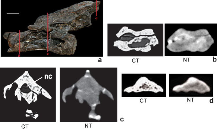
Figure 4. Axis (No. H25-1) of an undetermined diplodocid sauropod a) in left lateral view, red bars indicate axial cross-sections displayed in Figure 4b-d, scale bar (white) is 20 mm; b) axial section through the condyle: in the CT image (left) two large cavities filled with sediment and several smaller foramina are visible whereas in the NT image (right) even the large cavities are difficult to trace; c) axial section through the region of the diapophysis: in the CT image (left) several cavities and foramina are visible whereas the glue (quick drying adhesive) within the fractures is displayed as black as the background, in the NT image (right) the glue is detectable by its bright colour, nc = neural canal; d) axial section through the caudal part of the spinal process, only in the CT image (left) details of internal hollow spaces in the bone are visible.