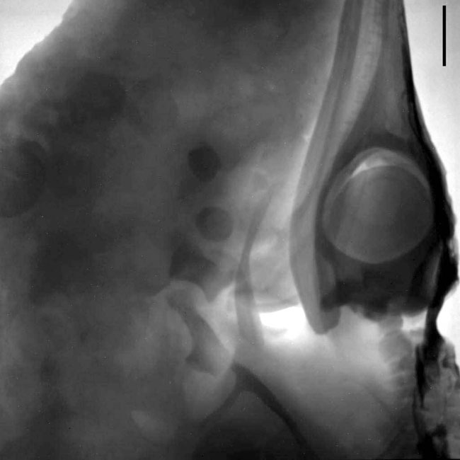
Figure 6. Neutron radiographic image of the head of an ichthyosaur imbedded in a shale. The image displays the orbital region of the skull with caudal part of the rostrum, orbit, braincase and first neck vertebrae (right side, from top to bottom) and some isolated bones of the forelimb (center of the image). The bone structure is clearly visible and can be distinguished from the pelitic matrix. This investigation was done on behalf of Urs Oberli (St. Gallen, Switzerland). Scale bar is 20 mm.