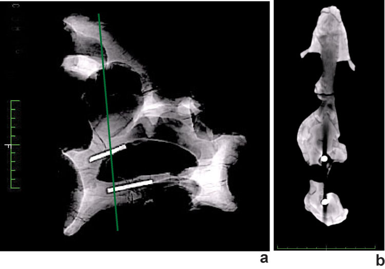
Figure 7. CT scan of a cross-section through the axis of Brachiosaurus brancai (SI 71) with two metal rods inside, analyzed by the Institute for Small Pets of the Free University in Berlin a) lateral overview about the vertebral with the location of the metal rods, b) transverse cross-section (position see green line in a), through axis showing the metal rods. The X-rays are strongly scattered, which considerably decreases the image quality. Green scale bar is 10 cm.