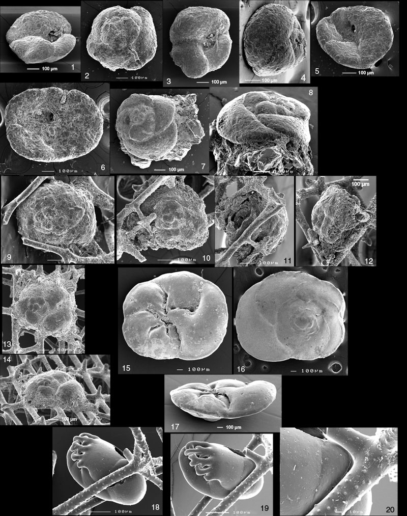
Figure 10.1-10.25. 1-3. cf. Tritaxis fusca Williamson. 1. Oblique-lateral view showing aperture. 2. Spiral side view (with small foreign tube attached). 3. Apertural side. Slurp gun sample SLRP4771, sponge fraction. 4-5. cf. Tritaxis fusca. 4. Oblique view of spiral side. 5. Oblique umbilical view showing aperture. Umbilicus filled with mud. Slurp gun sample SLRP4771, <1 mm. 6. cf. Tritaxis fusca. Apertural side of specimen still holding a spicule. There is some leftover material of the attachment cyst in the umbilical area. Slurp gun sample SLRP4771, <1 mm. 7-8. cf. Tritaxis fusca attached to fragment of Rhabdammina. 7. Spiral view. 8. Oblique view showing attachment cyst. Slurp gun sample SLRP4771, <1 mm. 9. cf. Tritaxis fusca attached to meshwork, spiral view. Slurp gun sample SLRP4771, <1 mm. 10-12. cf. Tritaxis fusca attached to meshwork. Stained attachment cyst. 10. Spiral view. 11. Side view from the left on Figure 10.10. 12. Side view from the right on Figure 10.10. Slurp gun sample SLRP4772, sponge fraction. 13-14. ?Tritaxis fusca attached to meshwork, with cyst. 13. Spiral view. 14. Oblique view. Shipek grab TUL99A017, sponge fraction. 15-17. Hyrrokkin cf. sarcophaga Cedhagen. 15. umbilical side. 16. spiral side. 17. edge view showing aperture. Shipek grab TUL99A015, >1 mm. 18-20. Nonionella digitata Nørvang. Trapped specimen. 18. the crack in the test along the spicule results from trying to remove the specimen with a brush. 19. different angle. 20. close-up of a boss that possibly developed in reaction to the presence of the spicule and seems to effectively hold the foraminifer in place. Piston core sample TUL99A09, 92-95 cm, >1 mm.