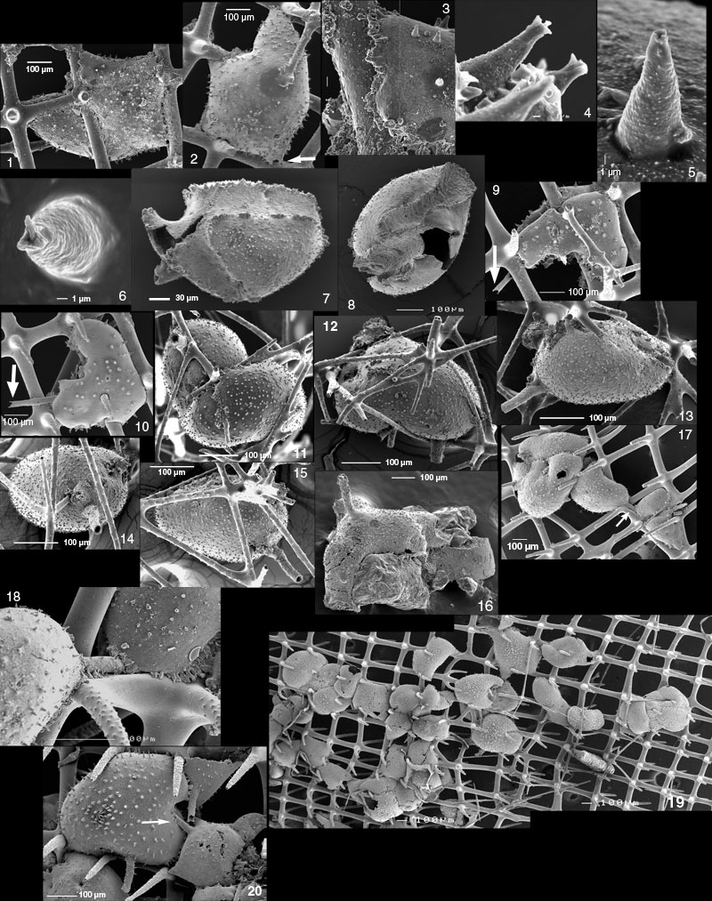
Figure 11.1-11.20. 1-6. GSC127659. Ramulina siphonifera n. sp. Attached (impaled) on Farrea occa meshwork. 1-2. opposite views, whole specimen. Arrow. aperture with exceptionally short siphon. 3. close-up of attachment to spicules, showing barbs or frills. 4. close-up of spines with bifurcating overgrowths at their tip. 5. close-up of spine without bifurcations. 6. close-up of spine with bifurcations. Shipek grab TUL99A014, sponge fraction. 7. GSC127660. Ramulina siphonifera fallen off its substrate, showing imprint of meshwork and frills at the limit between the outer wall and the spicule. The wall is spinose except for the part wrapping around the spicules. Shipek grab TUL99A015, sponge fraction. 8. GSC127661. Ramulina siphonifera showing imprint of sponge spicules. Piston core TUL99A09, 167-170 cm, <1 mm. 9-10. GSC127662. Ramulina siphonifera. Opposite sides of a specimen growing on F. occa. Arrow points at aperture. IKU grab TUL99A06 subcore, 6-9 cm depth, >1 mm including sponge fraction. 11-12. GSC127663. Ramulina siphonifera. Opposite views on specimen twisting inside the meshwork of Heterochone calyx. Piston sore TUL99A09, 167-170 cm depth, <1 mm. 13-15. GSC127658 (holotype) Ramulina siphonifera. Three different views. Piston core TUL99A09, 167-170 cm depth, <1 mm. 16. GSC127664. Ramulina siphonifera attached to sand grain. IKU grab TUL99A01, surface subsample ("forams" sample), sponge fraction. 17-18. GSC127665. Ramulina siphonifera. 17. Five specimens on F. occa. Arrow points at tube apparently joining two successive chambers. 18. Close-up of tube. frills around tube suggest that specimen at right came later and overlapped tube belonging to specimen at left. These are not successive chambers of the same specimen. IKU grab TUL99A01 subcore, 3-6 cm depth, >1 mm and sponge fraction combined. 19-20. GSC127666. Ramulina siphonifera. 19. Two-image composite showing R. siphonifera specimens clustering on F. occa meshwork. This view includes two specimens of Lobatula mckannai and one of Gaudryina accelerata (in the right hand part of the picture). 20. Close-up of a few specimens at extreme left of Figure 11.19. One apertural siphon is engulfed by a later specimen (arrow). IKU grab TUL99A01 subcore, 3-6 cm depth, >1 mm and sponge fraction combined.