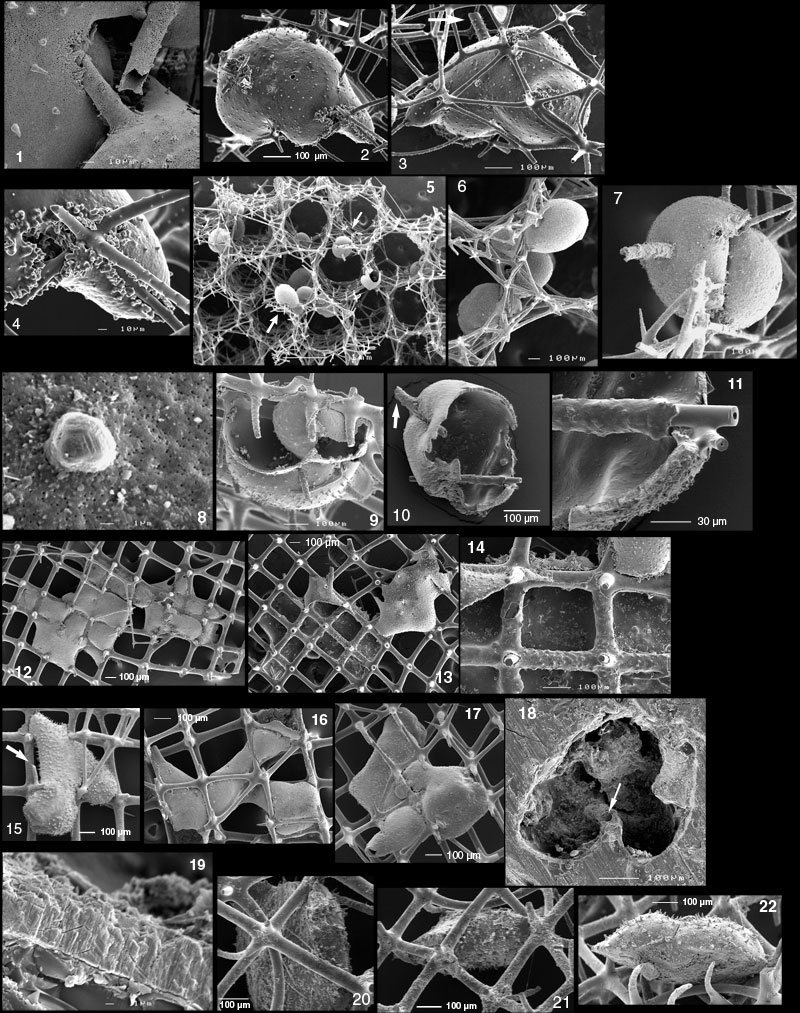
Figure 12.1-12.22. 1. GSC127666. Ramulina siphonifera n. sp. Same specimen as Figure 11.20, close-up of apertural siphon engulfed by later specimen. IKU grab TUL99A01 subcore, sample 3-6 cm, sponge fraction. 2-4. GSC127667. Ramulina siphonifera. Attached (impaled) inside Aphrocallistes vastus. 2-3. opposite sides, arrow points at aperture. 4. frills around sponge spicules. close-up of Figure 12.2. Piston core TUL99A09, sample 167-170 cm depth, >1 mm. 5-8. GSC127668. Many specimens of Ramulina siphonifera attached (impaled) on Aphrocallistes vastus. 5. low magnification view of specimens dispersed in meshwork. 6. group of specimens marked by lower arrow on Figure 12.5. 7. specimen marked by upper arrow on Figure 12.5. Where there is little constraining meshwork, R. siphonifera tends to assume a more or less spherical shape. 8. High magnification of the wall of the specimen of Figure 12.7. The large feature is the beginning of a spine. Some etching has taken place postmortem, hence the crystalline marks on the "spine." The pores may have been enlarged by dissolution. Shipek grab TUL99A019, sponge fraction. 9. GSC127669. Ramulina siphonifera. This species may be considered unilocular and this picture probably represents specimens engulfing each other, with broken-in walls. IKU grab TUL99A01 subcore, sample 3-6 cm, >1 mm. 10-11. GSC127670. Ramulina siphonifera. Broken in specimen, engulfing spicules. 10. general view, arrow points at apertural siphon, not to be confused with the spicule besides. 11. close-up of lower right part showing how the foraminifer wraps the engulfed spicules by a calcite wall. Shipek grab TUL99A017, sponge fraction. 12-14. GSC127671. Ramulina siphonifera. 12-13. opposite sides of one or more specimens growing on F. occa. All of the left part is just one chamber. On Figure 12.13, a large part of the wall of the left side is broken off, showing the interior of the opposite wall and the calcite layer wrapping the meshwork and insulating it from the protoplasm. 14. close-up of the meshwork and of the insulating calcite layer. A similar growth mode is reported in Thurammina from Jurassic sponge reefs. Shipek grab TUL99A014, sponge fraction. 15. GSC127672. Ramulina siphonifera on F. occa. Arrow points at aperture. Shipek grab TUL99A014, sponge fraction. 16. GSC127673. Ramulina siphonifera spreading through F. occa. Triggerweight core TUL99A010, sample 85-88, >1 mm. 17. GSC127674. Ramulina siphonifera spreading through F. occa. Triggerweight core TUL99A010, sample 85-88, >1 mm. 18-19. Ramulina siphonifera. 18. section through specimen embedded in Lakeside 70. Arrow points at a sponge spicule more or less normal to image plane. Note how wall wraps around the spicule. Compare with Figure 99 in Gaillard (1983). 19. close-up of wall showing radial structure. Interior of the test is up. Shipek grab TUL99A019, sponge fraction. 20-22. GSC127675. Ramulina siphonifera. Three specimens flattened on one side. All three attached to the same sponge fragment. The approximately flat side is non-spinose whereas the opposite side is spinose. These specimens may have grown on a soft substrate (sponge tissue?) which has not been preserved. Shipek grab TUL99A014, sponge fraction.