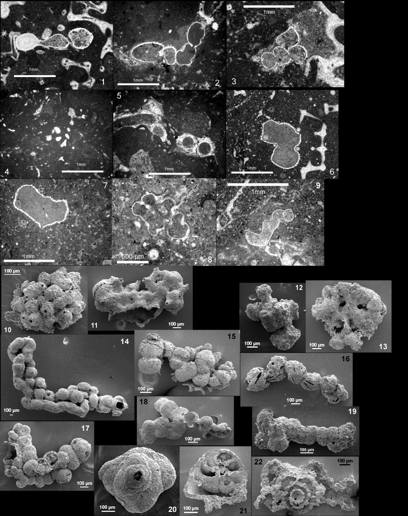
Figure 14.1-14.22. 1. Bullopora tuberculata (Sollas). Foraminifer laced-in within relatively coarse sponge meshwork. Tuejar near Chelva, Province Valencia, Spain, Oxfordian. 2. Bullopora tuberculata (Sollas). The lower and right edges of the picture are filled with sponge meshwork on which the Bullopora grows. See foramen. Hanner Steige, Urach, Swabian Alb, Germany, Uppermost Kimmeridgian. 3. Bullopora tuberculata (Sollas). Note foramina and central canal within spines. Closely associated with serpulids (on the right) and to microbial crust (left). Jabaloyas, Province Teruel, Spain, Oxfordian. 4. Vinelloidea crussolensis growing on sponge meshwork. Büchelberg near Urach, Swabian Alb, Germany, Uppermost Kimmeridgian. 5. Vinelloidea crussolensis (dark) grows around a serpulid (clear). Willmandingen, Swabian Alb, Germany, Early Kimmeridgian. 6. Thurammina papillata. Foraminifer is laced-in within coarse sponge meshwork. Jabaloyas, Province Teruel, Spain, Oxfordian.7. Thurammina papillata. The whole SE half of this picture is sponge meshwork to which the foraminifer is attached. Calatorao near La Almunia de Doña Godina, Province Zaragoza, Spain, Oxfordian. 8. Thurammina papillata. Foraminifer grows within sponge meshwork and is pierced and impaled by it. Spicule alignments cross the picture diagonally, at right angle to one another. Calatorao near La Almunia de Doña Godina, Province Zaragoza, Spain, Oxfordian. 9. Tolypammina vagans. Calatorao near La Almunia de Doña Godina, Province Zaragoza, Spain, Oxfordian. 10-22 are reillustrations of specimens figured in Schmalzriedt (1991). Specimens were loaned by the University of Tübingen and photographed with the SEM at Geological Institute in Stuttgart. 10-13. Thurammina papillata. 10. Specimen of Plate 1, fig. 7 in Schmalzriedt. 11. Plate 1, fig. 5. 12. Plate 1, fig. 6. 13. Plate 1, fig. 9. 10-11. apertures visible, not much evidence of meshwork presence. 12-13. imprint of meshwork clearly visible. 14. Tolypammina vagans. Plate 3, fig. 9 in Schmalzriedt. This specimen was growing through a now-dissolved meshwork whose presence left marks at many places. 15-19. Subbdelloidina hauesleri Frentzen. Schmalzriedt calls uniserial forms Placopsilina cenomana d'Orbigny and forms with irregular pile-ups of chambers (traubig), P. hauesleri (Frentzen). He names15, 16 and 18, P. cenomana; 17, P. hauesleri, and 19. P. cenomana/haeusleri transitional form. 15. Plate 5, fig. 10 in Schmalzriedt; imprint of spicules is visible. 16. Plate 5, fig. 8; rectilinear marks are left by spicules agglutinated by test and subsequently dissolved. 17. Plate 5, fig. 12, few spicule marks. 18. Plate 5, fig. 9; branching specimen. 19. Plate 5, fig. 14; partly uniserial and partly traubig. 20-22. Tritaxis lobata (Seibold and Seibold). 20. Plate 6, fig.10 in Schmalzriedt; spiral side. 21. Plate 6, fig.11; specimen broken along the vertical axis. 22. Plate 6, fig.22; horizontal cross-section including attachment cyst.