|
|
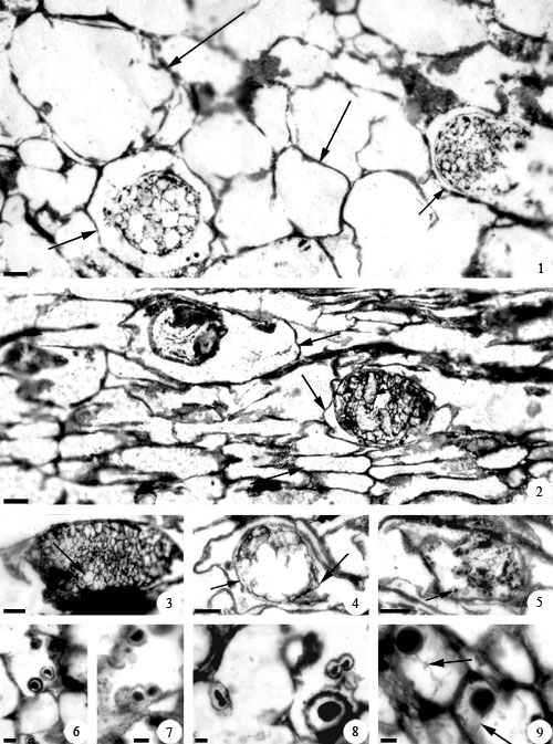
Figure 2. Synchytrium permicus sp. nov., silicified endoparasitic chytridiomycete from Skaar Ridge, Central Transantarctic Mountains, Antarctica. 1. Intact nearly spherical thin-walled sporangium and ruptured thin-walled sporangium, each within an enlarged host cell (lower arrows). Note polygonal segments inside sporangia. Middle arrow shows a normal size host cell. Upper arrow shows an empty enlarged host cell. Slide #21644, 11653 C-top, peel #1. Scale bar= 25 µm. 2. Elliptical thin-walled sporangium (right) within an enlarged host cell (middle left arrow) and thick-walled sporangium (left) within an enlarged host cell (upper arrow). Middle right arrow points to one of the transversely arranged globose segments that partially fill the lumen of the thin-walled sporangium. Lower arrow shows normal size host cell. Slide #21644, 11653 C-top, peel #1. Scale bar= 25 µm. 3. Elliptical thin-walled sporangium within an enlarged host cell. Oblique arrow indicates smaller globose segments that completely fill the lumen of the sporangium. Slide #21644, 11653 C-top, peel #1. Scale bar= 25 µm. 4. Broadly elliptical thin-walled sporangium partially filled with sporangial contents within an enlarged host cell. Left arrow indicates a single-layered sporangial wall. Right arrow shows a papilla-like projection on the sporangial wall. Slide #21644, 11653 C-top, peel #1. Scale bar= 25 µm. 5. Ruptured thin-walled sporangium within a host cell. Arrow shows zoospore within sporangium. Slide #21644, 11653 C-top, peel #1. Scale bar= 25 µm. 6. Pair of zoospores close to each other. Slide #21643, 11665 C-top, peel #1. Scale bar= 7.5 µm. 7. Pair of zoospores closely appressed to each other with distinct margins. Slide #21643, 11665 C-top, peel #1. Scale bar= 7.5 µm. 8. Pairs of zoospores attached to each other and with a common margin. Upper right: pair of zoospores with individual opaque bodies still distinct from each other. Left: pair of zoospores with fused opaque central bodies. Lower right: enlarged zoospore (zygote). Slide #21643, 11665 C-top, peel #1. Scale bar= 7.5 µm. 9. Enlarged zoospores (zygotes) in host tissue. Black arrows indicate thin flagellum at the base of each zoospore (zygote). Slide #21639, 11657 E-Top, peel #5. Scale bar= 7.5 µm.
|