|
|
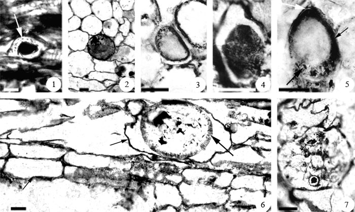
Figure 3. Synchytrium permicus sp. nov., silicified endoparasitic chytridiomycete from Skaar Ridge, Central Transantarctic Mountains, Antarctica. 1. Large zoospore within host tissue. Arrow indicates opaque inclusions in the lumen around the opaque central body. Slide #21648, 11654 F-top, peel #1β. Scale bar= 15 µm. 2. Large zoospore (possibly zygote or developing thick-walled sporangium) within host tissue. Note lumen completely filled with opaque particles. Slide #21639, 11657 E-Top, peel #5. Scale bar= 15 µm. 3. Curved-ellipsoid thick-walled sporangium within host cell. Slide #21643, 11665 C-top, peel #1. Scale bar= 25 µm. 4. Ovoid thick-walled sporangium within distorted host cell. Note pitted outer surface. Slide #21643, 11665 C-top, peel #1. Scale bar= 25 µm. 5. Pyriform thick-walled sporangium within enlarged host cell. Right arrow indicates sporangial wall. Upper arrow shows a small slit at sporangium end. Lower arrow shows zoospore within sporangium. Slide #21643, 11665 C-top, peel #1. Scale bar= 25 µm. 6. Ellipsoid thick-walled sporangium within enlarged host cell (middle arrow). Left arrow indicates normal size cell. Right arrow show ridges at the perimeter of the sporangium. Slide #21644, 11653 C-top, peel #1. Scale bar= 25 µm. 7. Ruptured thick-walled sporangium. Note zoospore within sporangium. Slide #21644, 11653 C-top, peel #1. Scale bar= 25 µm.
|