|
|
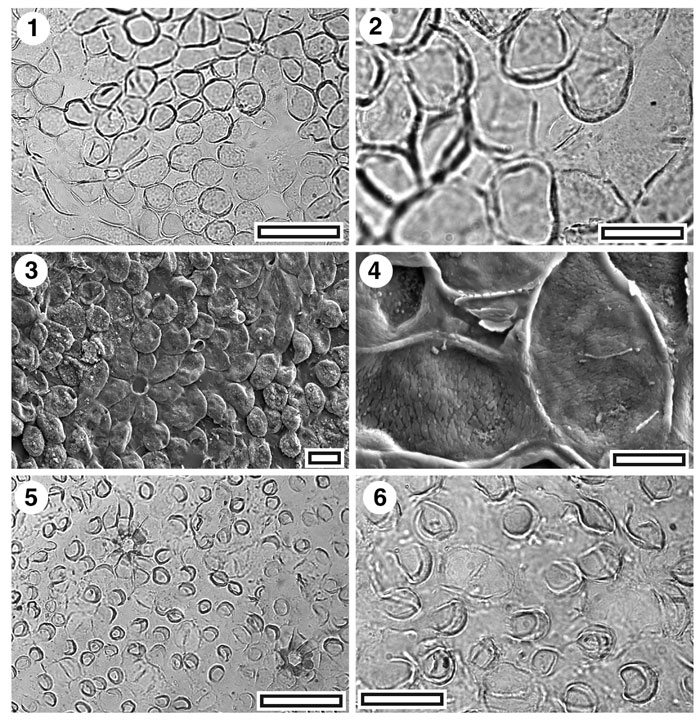
Figure 12. Lauraceae. CUT-L-JEC, and CUT-L-GCI. 1. CUT-L-JEC. TLM view showing papillae, and (upper right) a trichome insertion scar with a distinct ring of foot cells (SB0719, scale-bar = 50 µm); 2. CUT-L-JEC. TLM detail of a single stomatal complex (right of centre) surrounded by papillae (SB0719, scale-bar = 20 µm); 3. SEM view of outer cuticular surface showing trichome insertion scar with prominent foot cells (left of centre) and papillae obscuring stomata (S-1684, scale bar = 20 µm); 4. SEM view of inner cuticular surface showing a possible stomatal complex (just above centre) (S-1684, scale-bar = 10 µm); 5. CUT-L-GCI. TLM view showing papillae and two trichome insertion scars with prominent thickening around the pores (SL5372, scale-bar = 50 µm); 6. CUT-L-GCI. TLM detail with a stomatal complex visible (centre left) SL5372, scale-bar = 20 µm).
|