|
|
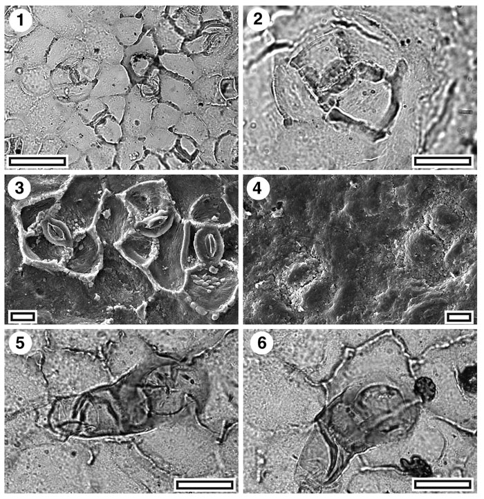
Figure 13. Lauraceae. CUT-L-DCG, 1. TLM view showing five stomatal complexes around a trichome (to right of centre) inserted over two epidermal cells (SB0346, scale-bar = 50 µm); 2. TLM detail of a single stomatal complex. Note massive thickening around stomatal pore and subsidiary cells (SB0346, scale-bar = 20 µm); 3. SEM view of inner cuticular surface showing three stomatal complexes. Note prominent thickening of subsidiary cell walls (S-1544, scale-bar = 10 µm); 4. SEM view of outer cuticular surface showing two stomatal complexes. Subsidiary cells are visible and the stomatal pore is slit-like and largely plugged (S-1544, scale-bar = 10 µm); 5. TLM view of a trichome over junction of two epidermal cells (SL5049, scale-bar = 20 µm); 6. TLM view of a trichome over junction of two epidermal cells. Note epiphyllous germling (SL5049, scale-bar = 20 µm).
|