|
|
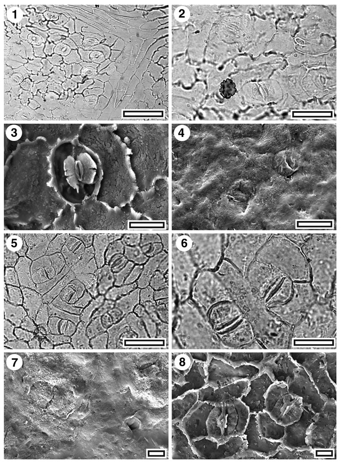
Figure 14. Lauraceae. CUT-L-DCD and CUT-L-DCI. 1. CUT-L-DCD. TLM view showing several stomatal complexes and (upper right) the junction of two fine veins indicated by distinct venal epidermal cells (SB0349, scale-bar = 50 µm); 2. CUT-L-DCD. TLM detail of a four stomatal complexes (note epiphyllous germling at lower left) (SB0349, scale-bar = 20 µm); 3. UT-L-DCD. SEM view of inner cuticular surface showing a single stomatal complex. Note "butterfly" cuticular scales (S-1683, scale-bar = 10 µm); 4. CUT-L-DCD. SEM view of outer cuticular surface showing two stomatal complexes, barely distinguished from the surrounding topography (S-1683, scale-bar = 10 µm); 5. CUT-L-DCI. TLM view showing several stomatal complexes and (lower left) a trichome insertion scar. Note distinctive "anisocytic' pattern around the complexes (SB0672, scale-bar = 50 µm); 6. CUT-L-DCI. TLM detail of a two stomatal complexes (SB0672, scale-bar = 20 µm); 7. CUT-L-DCI. SEM view of outer cuticular surface showing two stomatal complexes (left of centre and upper right) where the subsidiary cells are visible and the pore slit-like, and a trichome insertion scar (lower right) (S-0302, scale-bar = 10 µm); 8. CUT-L-DCI. SEM view of inner cuticular surface showing two stomatal complexes (S-0302, scale-bar = 10 µm).
|