|
|
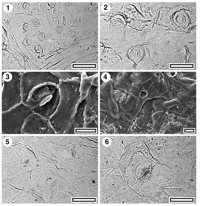
Figure 17. Lauraceae. CUT-L-JBI and CUT-L-JBA, 1. CUT-L-JBI. TLM view showing several stomatal complexes and (lower left) a trichome insertion scar over venal epidermal cells (SB0700, scale-bar = 50 µm); 2. CUT-L-JBI. TLM detail of three stomatal complexes. Note cuticular thickening surrounding complexes (SB0700, scale-bar = 20 µm); 3. SEM view of inner cuticular surface showing single stomatal complex. Note "butterfly" cuticular scales (S-1538, scale bar = 10 µm); 4. SEM view of outer cuticular surface showing two stomatal complexes (centre left and lower right) and two trichome insertion scars (centre and upper right) (S-1538, scale-bar = 10 µm); 5. CUT-L-JBA. TLM view showing three stomatal complexes (SB0391, scale-bar = 50 µm); 6. CUT-L-JBA. TLM detail of a single stomatal complex (SB0391, scale-bar = 20 µm).
|