|
|
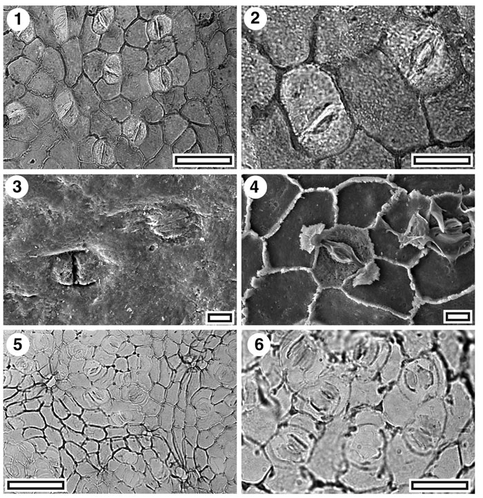
Figure 18. Lauraceae. CUT-L-GCG and CUT-L-GCH. 1. CUT-L-GCG. TLM view showing several stomatal complexes. Note typically thinner cuticle over subsidiary cells (SL1023, scale-bar = 50 µm); 2. CUT-L-GCG. TLM detail of two stomatal complexes (SL1023, scale-bar = 20 µm); 3. SEM view of outer cuticular surface showing two stomatal complexes with slit-like pores (S-1690, scale-bar = 10 µm); 4. SEM view of inner cuticular surface showing two stomatal complexes. The central specimen clearly shows the triangular flaps of cuticle at the polar ends of the complex (S-1690, scale-bar = 10 µm); 5. TLM view showing several stomatal complexes and three trichome insertion scars over venal epidermal cells (SL5374, scale-bar = 50 µm); 6. TLM detail of a group of stomatal complexes. Note very thin cuticle of the subsidiary cells, and clear "butterfly" shaped cuticular scales (SL5374, scale-bar = 20 µm).
|