|
|
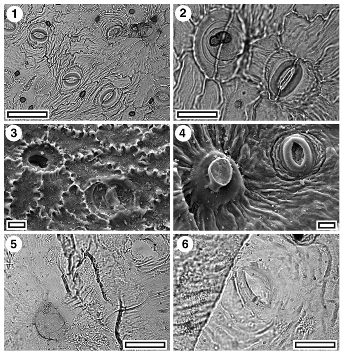
Figure 19. Proteaceae. CUT-P-EJG and CUT-P-EJF. 1. CUT-P-EJG. TLM view showing several stomatal complexes and (upper right) a persistent trichome (SB0674, scale-bar = 50 µm); 2. CUT-P-EJG. TLM detail of a trichome base over two epidermal cells (left) and a stomatal complex (right) (SB0674, scale-bar = 20 µm); 3. CUT-P-EJG. SEM view of inner cuticular surface showing a trichome base (upper left) and a stomatal complex (lower right) (S-1696, scale-bar = 10 µm); 4. CUT-P-EJG. SEM view of outer cuticular surface showing a trichome base (centre left) and a stomatal complex (upper right) (S-1696, scale-bar = 10 µm); 5. CUT-P-EJF. TLM view showing a trichome base (lower left) and (upper right) a stomatal complex with the guard cell cuticle broken away (SB0679, scale-bar = 50 µm); 6. CUT-P-EJF. TLM detail of a single stomatal complex (SB0679, scale-bar = 20 µm).
|