|
|
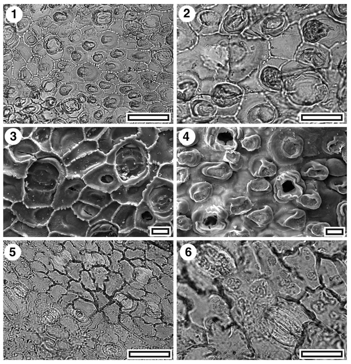
Figure 21. Proteaceae. CUT-P-GDJ and CUT-P-EAA. 1. CUT-P NEW. TLM view showing several stomatal complexes (SL5412, scale-bar = 50 µm); 2. CUT-P NEW. TLM detail of a trichome base over two epidermal cells (left of centre), a stomatal complex (lower right). Other dark objects are mostly papillae (SL5412, scale-bar = 20 µm); 3. SEM view of inner cuticular surface showing four stomatal complexes, and papillate epidermal cells (S-1698, scale-bar = 10 µm); 4. SEM view of outer cuticular surface showing three stomatal complexes (note the irregular ridges surrounding them), papillate epidermal cells, and (upper right) a trichome base (S-1698, scale-bar = 10 µm); 5. CUT-P-EAA. TLM view showing several stomatal complexes (SB0688, scale-bar = 50 µm); 6. CUT-P-EAA. TLM detail of a two stomatal complexes (SB0688, scale-bar = 20 µm).
|