|
|
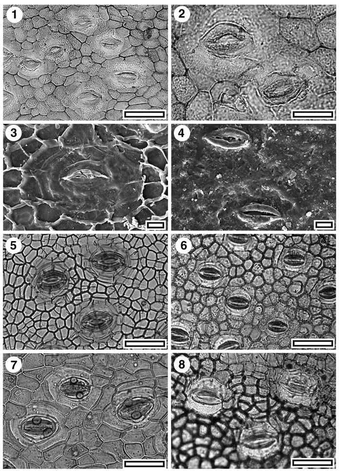
Figure 24. Rhizophoraceae, fossil and extant. 1. CUT-Z-JAG. TLM view showing several stomatal complexes (note alignment and relatively small epidermal cells) SL5251, scale-bar = 50 µm); 2. CUT-Z-JAG. TLM detail of two stomatal complexes (SL5251, scale-bar = 20 µm); 3. CUT-Z-JAG. SEM view of inner cuticular surface showing single stomatal complex. Note most of subsidiary cell walls have collapsed (S-1545, scale-bar = 10 µm); 4. CUT-Z-JAG. SEM view of outer cuticular surface showing two stomatal complexes. Note prominent elliptical outer stomatal ledges (S-1545, scale-bar = 10 µm); 5. Extant Rhizophora mangle., TLM view showing three stomatal complexes (note alignment and relatively small epidermal cells) OPH3063, scale-bar = 50 µm); 6. Extant Bruguiera sexangula, TLM view showing several stomatal complexes. Note alignment and relatively small epidermal cells (CANB 231858, scale-bar = 50 µm); 7. Extant Pelliciera rhizophorae, TLM view showing three stomatal complexes. Note alignment and relatively small epidermal cells (OPH7495, scale-bar = 50 µm); 8. Extant Ceriops tagal, TLM view showing three stomatal complexes. Note alignment and relatively small epidermal cells (AQ109064, scale-bar = 50 µm).
|