|
|
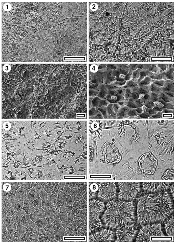
Figure 28. CUT-Z-JBB, 1. TLM view showing three areoles with papillae-covered cells separated by venal epidermal cells (SL5371, scale-bar = 50 µm); 2. TLM detail of papillae, with completely obscured stomatal complexes (SL5371, scale-bar = 20 µm); 3. SEM view of outer cuticular surface showing papillae entirely obscuring stomatal complexes (S-1699, x600 scale-bar = 20 µm); 4. SEM view of inner cuticular surface showing five deeply sunken stomata (S-1699, scale-bar = 10 µm); 5. TLM view showing widely-spaced papillae (SB0378, scale-bar = 50 µm); 6. TLM detail of widely-spaced papillae (SB0378, scale-bar = 20 µm); 7. TLM view showing closely-spaced papillae (SB0710, scale-bar = 50 µm); 8. TLM detail of closely spaced papillae (SB0710, scale-bar = 20 µm).
|