|
|
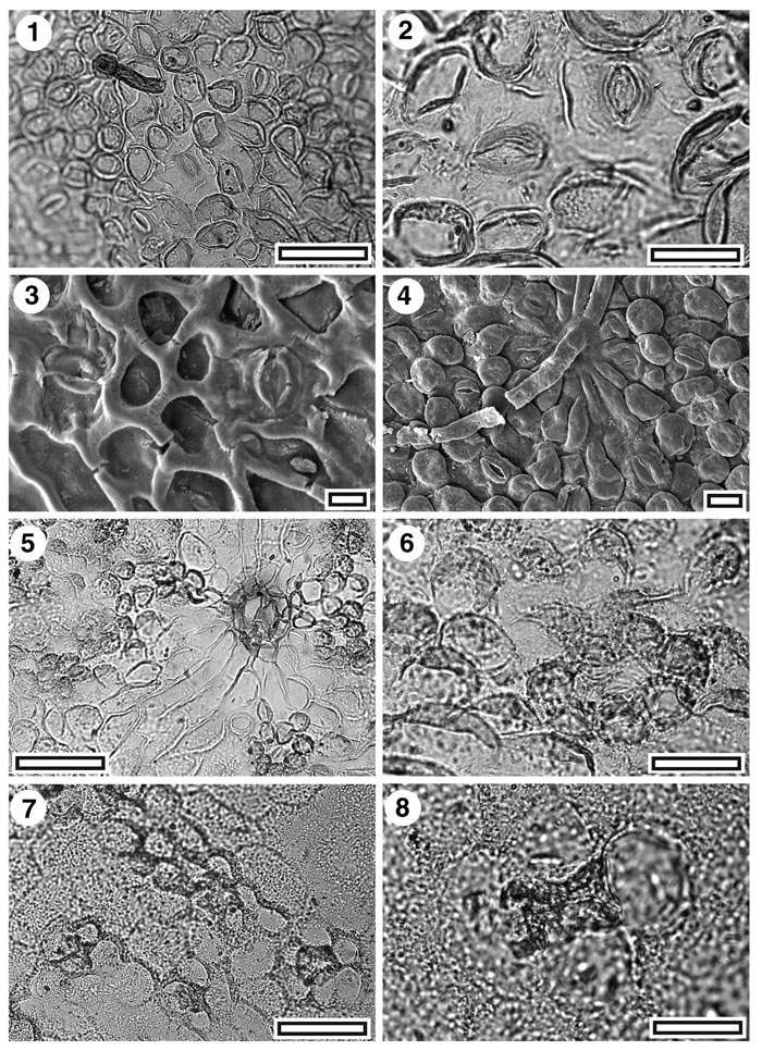
Figure 29. CUT-Z-GDA, CUT-Z-Z-GCF, and CUT-Z-Z-JAD, 1. CUT-Z-GDA. TLM view showing scattered stomatal complexes and (upper left) a darkly staining persistent trichome (SL5433, scale-bar = 50 µm); 2. CUT-Z-GDA. TLM detail of two stomatal complexes (SL5433, scale-bar = 20 µm); 3. CUT-Z-GDA. SEM view of inner cuticular surface showing two stomatal complexes (S-1688, scale-bar = 10 µm); 4. CUT-Z-GDA. SEM view of outer cuticular surface showing persistent trichome (upper centre) and several stomatal complexes surrounded by papillate epidermal cells (S-1688, scale-bar = 10 µm); 5. CUT-Z-GCF. TLM view showing clusters of papillae surrounding stomatal complexes and a large trichome insertion scar in the middle of venal epidermal cells (SB0371, scale-bar = 50 µm); 6. CUT-Z-GCF. TLM detail showing a ring of papillae around a stomatal complex (lower right) (SB0371, scale-bar = 20 µm); 7. CUT-Z-JAD. TLM view showing several stomatal complexes surrounded by subdued papillae (SB0725, scale-bar = 50 µm); 8. CUT-Z-JAD. TLM detail of a single stomatal complex. Note massive thickening around pore (SB0725, scale-bar = 20 µm).
|