|
|
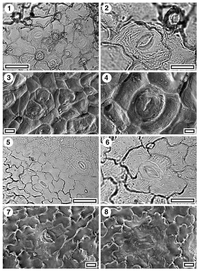
Figure 31. CUT-Z-JCC and CUT-Z-Z-JJA, 1. CUT-Z-JCC. TLM view showing several stomatal complexes and persistent trichomes at intersections of epidermal cells (SB0741, scale-bar = 50 µm); 2. CUT-Z-JCC. TLM detail of a single stomatal complex (note ornamentation of fine ridges on the subsidiary cells) and (upper right) a trichome (SB0741, scale-bar = 20 µm); 3. SEM view of inner cuticular surface showing several stomatal complexes (S-0328, x600 scale-bar = 20 µm; 4. SEM view of inner cuticular surface showing a single stomatal complex (S-0328, scale-bar = 10 µm); 5. CUT-Z-JJA. TLM view showing three stomatal complexes (SB0723, scale-bar = 50 µm); 6. CUT-Z-JJA. TLM detail of a single stomatal complex (SB0723, scale-bar = 20 µm); 7. CUT-Z-JJA. SEM view of inner cuticular surface showing a single stomatal complex (S-0313, scale-bar = 10 µm); 8. CUT-Z-JJA. SEM view of inner cuticular surface showing a single stomatal complex (S-0313, scale-bar = 10 µm).
|