|
|
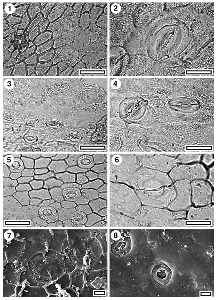
Figure 32. CUT-Z-JEB, CUT-Z-Z-JEJ, and CUT-Z-Z-JEA, 1. CUT-Z-JEB. TLM view showing three stomatal complexes and (centre left) a massively thickened trichome insertion scar (SB0753, scale-bar = 50 µm); 2. CUT-Z-JEB. TLM detail of a single stomatal complex (SB0753, scale-bar = 20 µm); 3. CUT-Z-JEJ. TLM view showing several stomatal complexes (SB0726, scale-bar = 50 µm); 4. CUT-Z-JEJ. TLM detail of two stomatal complexes (SB0726, scale-bar = 20 µm); 5. CUT-Z-JEA. TLM view showing several stomatal complexes (SB0717, scale-bar = 50 µm); 6. CUT-Z-JEA. TLM detail of a single stomatal complex (SB0717, scale-bar = 20 µm); 7. CUT-Z-JEA. SEM view of inner cuticular surface showing two stomatal complexes (S-1541, scale bar = 10 µm); 8. CUT-Z-JEA. SEM view of outer cuticular surface showing two stomatal complexes. Note very circular outer stomatal ledge (S-1541, scale-bar = 10 µm).
|