|
|
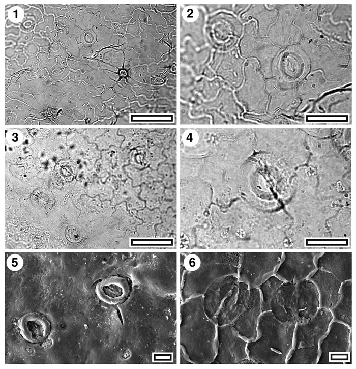
Figure 35. CUT-Z-JCJ and CUT-Z-Z-JJH, 1. CUT-Z-JCJ. TLM view showing several stomatal complexes and two trichome attachment scars (SB0763, scale-bar = 50 µm); 2. CUT-Z-JCJ. TLM detail of two stomatal complexes (SB0763, scale-bar = 20 µm); 3. CUT-Z-JJH. TLM view showing TLM view showing four stomatal complexes. Note the pair to left of centre is networked - sharing a single contact cell (SB0701, scale-bar = 50 µm); 4. CUT-Z-JJH. TLM detail of a single stomatal complex (SB0701, scale-bar = 20 µm); 5. CUT-Z-JJH. SEM view of outer cuticular surface showing two stomatal complexes (S-1689, scale-bar = 10 µm); 6. CUT-Z-JJH. SEM view of outer cuticular surface showing two stomatal complexes (S-1689, scale-bar = 10 µm).
|