|
|
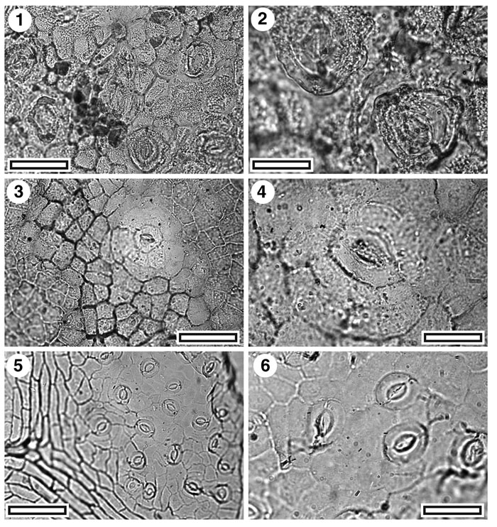
Figure 36. CUT-Z-JDH, CUT-Z-Z-JAA, and CUT-Z-Z-JJI, 1. CUT-Z-JDH. TLM view showing several stomatal complexes (SB0712, scale-bar = 50 µm); 2. CUT-Z-JDH. TLM detail of two stomatal complexes. Note massive rings of cuticle surrounding each. (SB0712, scale-bar = 20 µm); 3. CUT-Z-JAA. TLM view showing a single stomatal complex (SB0350, scale-bar = 50 µm); 4. CUT-Z-JAA. TLM detail of a single stomatal complex (SB0350, scale-bar = 20 µm); 5. CUT-Z-JJI. TLM view showing several stomatal complexes and (at left) a vein reflected in the shape of venal epidermal cells (SB0754, scale-bar = 50 µm); 6. CUT-Z-JJI. TLM detail of four stomatal complexes (SB0754, scale-bar = 20 µm).
|