|
|
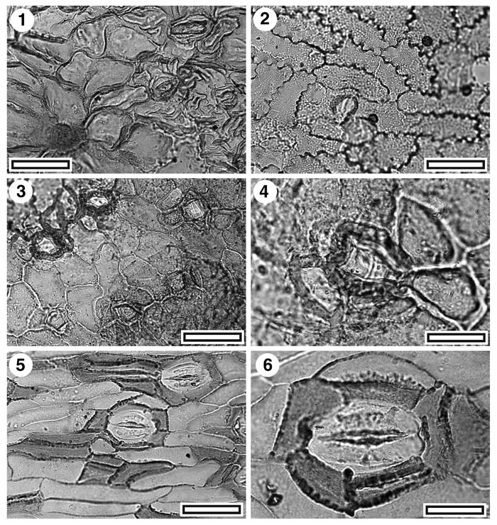
Figure 4. Pterostoma sp. and Bowenia sp. 1. Pterostoma hirsurtus. TLM view showing stomata at right, surrounded by prominent ridges of cuticle, and, lower left, a trichome base (SL5381, scale-bar = 50 µm); 2. ?Pterostoma. TLM view showing a single stomatal complex (SB0384, scale-bar = 50 µm); 3. ?Pterostoma. TLM view showing five stomatal complexes (SB0690, scale-bar = 50 µm); 4. ?Pterostoma. TLM detail of a single stomatal complex. Note massively thickened ring around stomatal pore (SB0690, scale-bar = 20 µm); 5. Bowenia. TLM view showing two stomatal complexes. Note differently thickened peristomatal walls of epidermal and subsidiary cells (SL5059, scale-bar = 50 µm); 6. CUT-Z-ACB. TLM detail of a single stomatal complex (SL5335, scale-bar = 20 µm).
|