|
|
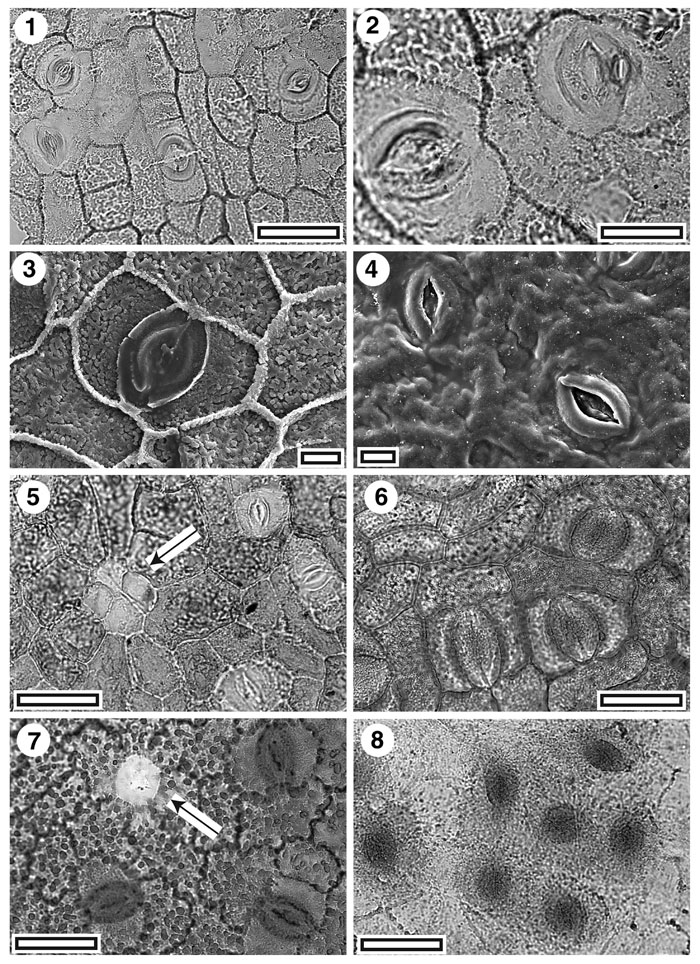
Figure 8. Winterceae, fossil and extant. 1. CUT-L-DDD. TLM view showing stomatal complexes (SB0756, scale-bar = 50 µm); 2. CUT-L-DDD. TLM view showing two stomatal complexes (SB0756, scale-bar = 20 µm); 3. SEM view of inner cuticular surface showing single stomatal complex. Note very granular surface of epidermal cells (S-1537, scale bar = 10 µm); 4. SEM view of outer cuticular surface showing two stomatal complexes. Note prominent outer stomatal ledges, but without plugged pores (S-1537, scale-bar = 10 µm); 5. CUT-L-DDD. TLM view showing lid cell complex indicated with an arrow (SB0360, scale-bar = 50 µm); 6. Extant Zygogynum balansae, TLM view showing three stomatal complexes. (AQ391241, scale-bar = 50 µm); 7. Extant Bubbia semecarpoides, TLM view showing lid cell indicated with an arrow (AQ546547, scale-bar = 50 µm); 8. Extant Belliolum burttianum, TLM view showing a group of stomatal complexes. (AQ463392, scale-bar = 50 µm).
|