|
|
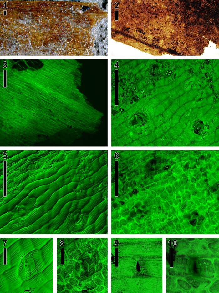
Figure 13. Cuticle morphotype 1, probable monocot, USNM 535049. (1) Strap-shaped cuticle viewed directly on rock (note adhering sand grains) under stereomicroscope, showing margin at top, broken at right; cells visible. (2) Light micrograph of mounted cuticle, margin at bottom, broken at right. (3) Cuticle under light microscope with epifluorescence showing elongate epidermal cells; square hypodermal cells at bottom. (4) Maximum brightness projection view of stacked confocal images, showing rectangular epidermal cells and sunken paracytic stomata. Note striated pattern on subsidiary cells. (5) Three-dimensional surface rendering of stacked confocal images, showing epidermal cells with striated surfaces and sunken stomatal areas. (6) Sum projection view of stacked confocal images, showing faint striated pattern from epidermal cells, thick-walled, square hypodermal cells, and stomatal areas. (7) Three-dimensional surface rendering of stacked confocal images showing epidermal cells with striated surface and a single stomatal area. (8) Sum projection view of previous figure showing guard cells, thick-walled hypodermal cells, and striated subsidiary cells. (9) Three-dimensional surface rendering of stacked confocal images, showing epidermal cells with striated surfaces, and a single stomatal area. (10) Sum projection view of previous figure showing guard cells, thick-walled hypodermal cells, and striated subsidiary cells. (1-6): Scale bars equal 100 µm; (7-10): Scale bars equal 30 µm.
|