|
|
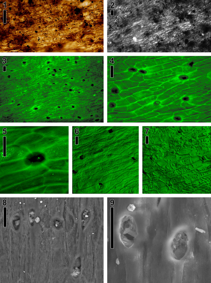
Figure 14. Cuticle morphotype 2, probable liverwort, USNM 535888. (1) Light micrograph of mounted cuticle; cells elongate, containing brown to black organic matter and minerals. (2) Sum projection view of stacked differential interference contrast images (light microscope); view similar to (1) but minerals and sediment more prominent. (3) Extended depth-of-focus image of stacked epifluorescence light micrographs; epidermal cells elongate and bearing pores. (4) Higher magnification, extended depth-of-focus image of stacked epifluorescence light micrographs; epidermal cells elongate and bearing pores with raised margins. (5) Enlarged sum projection view of stacked confocal images. The end walls between epidermal cells are visible to the bottom of the single raised pore. Note the brightly fluorescing pyrite in pore and in cells adjacent to pore. (6) Three-dimensional surface rendering of stacked confocal images, showing epidermal cells and open pores. (7) Same rendering as in (6), showing reverse side. Note minerals (rough 'bumps') and protruding epidermal end walls. All scale bars equal 20 µm.
|