|
|
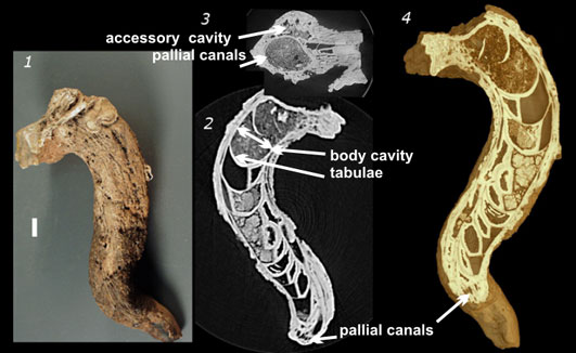
FIGURE 1. Caprinuloidea perfecta Palmer, 1928, NPL4387. 1.1, Complete specimen of RV-AV capped by debris. 1.2, Digital slice through the lower right valve (RV-AV) showing concave growth tabulae in the main body cavity, and accessory cavity following the line of the main cavity. Thin tubular pallial canals line the outer walls. 1.3, Digital cross-section through the right valve showing body cavity and accessory cavity. 1.4, Three-dimensional reconstruction with virtual cutaway revealing internal details, colored to approximate actual fossil specimen. The slice is the same orientation as the image of the specimen. Scale bar on 1.1 represents 1 cm and is valid for 1.2 and 1.3.
|