|
|
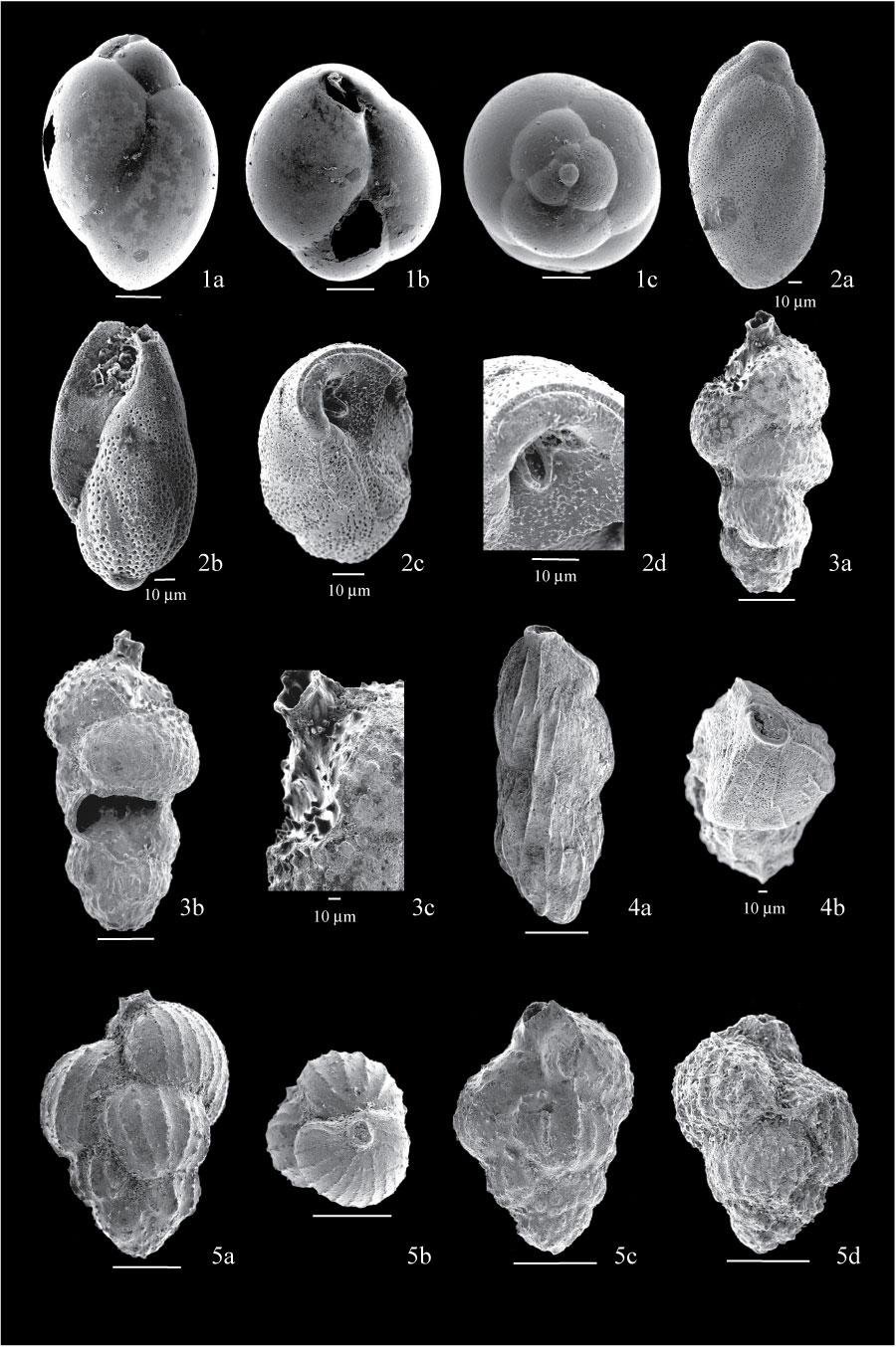
Figure 10. 1. Protoglobobulimina pupoides, three specimens from sample FC04-76; 1a, side view showing strongly overlapping chambers; 1b, apertural view showing internal toothplate; 1c, basal view showing circular cross-section and triserial arrangement. 2. Buliminella elegantissima; 2a, side view of specimen from sample BE1, showing close spiral; 2b, side view of specimen from sample FC04-64, showing aperture obscured by debris; 2c, apertural view of specimen from sample FC04-63, showing internal toothplate; 2d, detail of aperture of specimen 2c. 3. Euuvigerina aculeata from sample FC04-52; 3a, side view showing elongated shape; 3b, side view showing etched surface, spines uniting to form ribs at the base of test can be discerned; 3c, detail of tubular neck, showing remnants of cone-shaped spines at the base. 4. Angulogerina fluens Todd, from sample BE1; 4a, side view showing elongate test and longitudinal costae crossing the sutures; 4b, apertural view showing trigonal cross-section. 5. Euuvigerina peregrina; 5a, side view of specimen from sample FC04-53 showing clear longitudinal costae; 5b, apertural view of specimen 5a showing rounded aperture on top of tubular neck; 5c, side view of specimen from sample FC04-55, showing etched surface; 5d, side view of specimen from sample FC04-52 showing less developed costae and well developed pustules between them. All scales are 100 µm unless otherwise indicated.
|