|
|
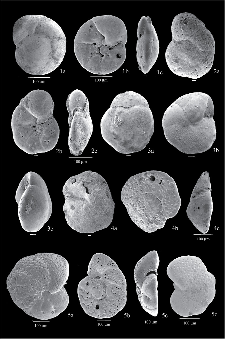
Figure 11. 1. Gavelinopsis campanulata from sample BE1; 1a, dorsal view; 1b, ventral view showing umbilical plug; 1c, side view showing more convex dorsal side. 2. Rosalina columbiensis from sample BE1; 2a, dorsal side showing coarse pores; 2b, ventral view showing aperture extending back along umbilicus; 2c, side view showing lipped marginal aperture. 3. Epistominella vitrea from sample BE2; 3a, dorsal side showing smooth test; 3b, ventral view showing radial sutures; 3c, side view. 4. Lobatula fletcheri from sample BE1; 4a, dorsal view showing lipped aperture; 4b, ventral view showing coarse perforation; 4c, side view showing well developed umbilical view on spiral side. 5. Lobatula lobatula from sample BE1; 5a, dorsal view showing very coarse perforation; 5b, ventral view showing aperture extending on the last few chambers; 5c, side view showing lipped aperture; 5d, dorsal view of another specimen with a more ovate outline. All scales are 10 µm unless otherwise indicated.
|