|
|
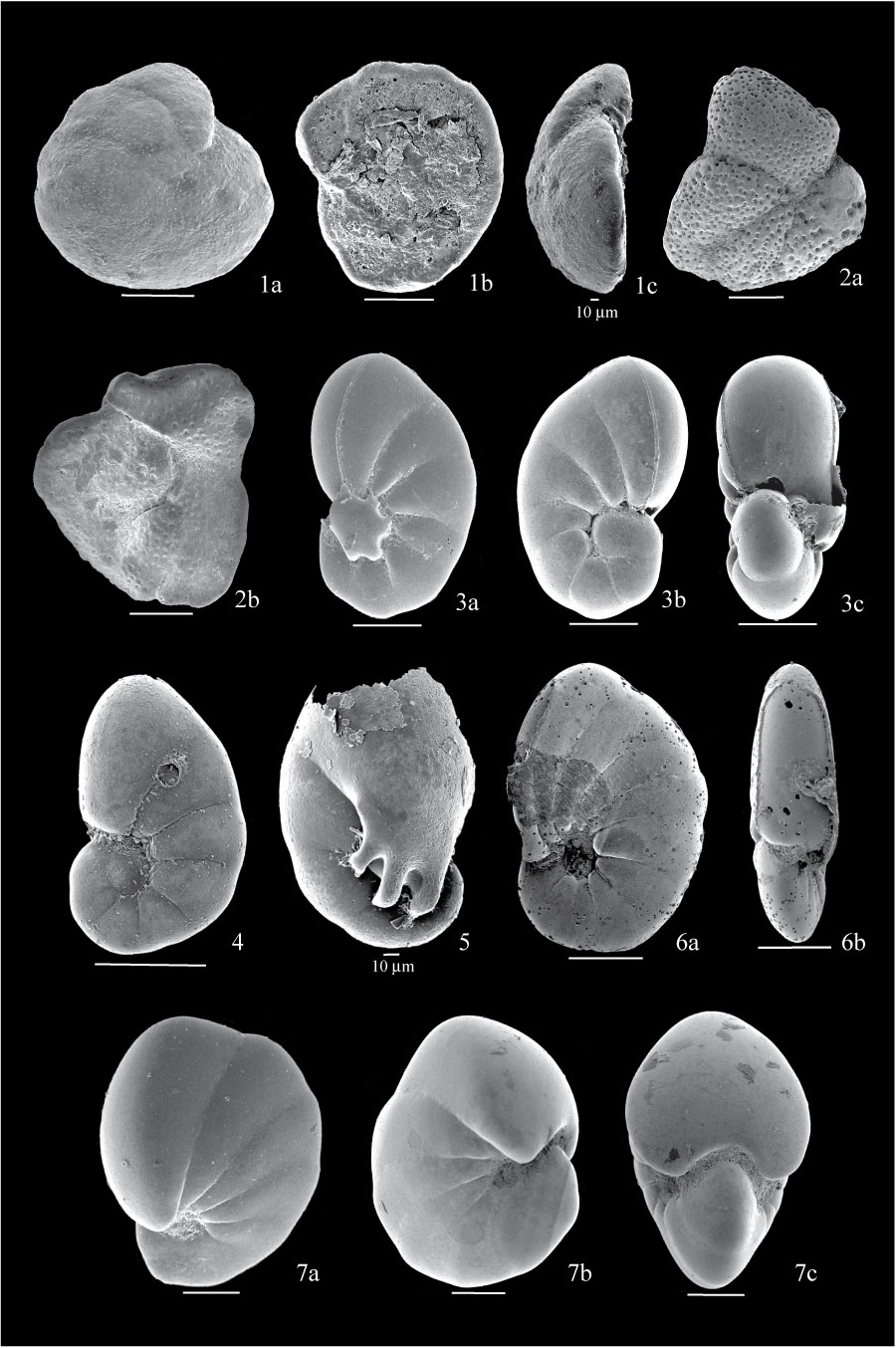
Figure 12. 1. Lobatula mckannai from sample BE1; 1a, dorsal view; 1b, ventral view covered with debris; 1c, side view showing aperture. 2. Dyocibicides biserialis from sample BE1; 2a, dorsal view showing irregular outline; 2b, ventral view showing coarse perforation. 3. Nonionella stella from sample BE1; 3a, side view showing umbilical flap extending from broken last chamber and covering the umbilicus; 3b, view of other side; 3c, apertural view showing basal arcuate aperture. 4. Nonionella cf. turgida from sample BE1, side view. 5. Nonionella digitata Nørvang from sample FC04-76, side view with broken last chamber, showing finger-like projections over umbilicus. 6. Pseudononion basispinata from sample BE2; 6a, side view showing hispid material in open umbilicus; 6b, apertural view showing compressed test. 7. Nonionellina labradorica from sample BE1; 7a, side view; 7b, view of the other side showing pustules covering umbilicus; 7c, apertural view showing broad flat apertural face. All scales are 100 µm unless otherwise indicated.
|